The success rate for labral tear hip surgery is high in most cases, however, depending on a variety of factors and the cause of the labral injury, a repeat procedure may be necessary after the primary surgery. One study found that 17% of patients required a second surgery.
In addition, physical activities after surgery in some cases are resumed normally, while in others are reduced to lower impact.
Labral Tear
Definition: Labral Tear
 The labrum is a tissue known as fibrocartilage that lines the rim of the hip socket. It helps to provide cushion, stability and depth to the socket creating protection of the joint and a seal for the joint.
Injury to the labrum can be caused by acute injury or chronic wear and tear of the joint. Sports such as soccer, hockey or ice hockey, ballet and golf display frequent causes of labral tear due to their repetitive motions of pivoting, twisting, kicking, running into other players or falling. Motor vehicle accidents or other type of trauma can also be a cause of labral tear. In addition bone structure abnormalities can also cause labral tear due to an abnormal wear and tear of the joint.
Treatment Options: Labral Tear
Non-Surgical: Labral Tear Hip
It is often better to try a non-surgical approach to treatment of a labral tear in the hip joint before surgery when possible. However, in some cases surgery may be recommended promptly. The labrum, due to its tough fibrocartilaginous make-up, has limited blood supply and cannot easily (if at all) repair itself or reattach itself to bone. For this reason, the long-term use of non-surgical treatments is discouraged and prompt manual repair is recommended.
There are a number of different non-surgical methods for treating labral tears and the pain and discomfort that accompany this type of injury.
- Physical Therapy- Physical Therapists can provide stretches, exercises and other therapeutic practices to help strengthen the hip joint and treat labral tears.
- Oral Medications- Oral, over the counter anti-inflammatory medications such as Tylenol or ibuprofen can be helpful, however, studies have begun to warn against the chronic or overuse of these types of medications (non-steroidal anti-inflammatory drugs NSAIDs) stating that they can make pain worse and lead to the eventual or accelerated need for joint replacement. Consult your physician before deciding to treat yourself with NSAIDs.
- Injections- Anesthetic and/or corticosteroids can be injected directly into the joint to provide relief from pain and inflammation. There has also been warnings against this type of treatment over the long term. Research has shown that frequent, long-term corticosteroid use can cause further degeneration and does not “heal” a tear.
- Rest- Rest from activities that cause pain to the hip is advised to help alleviate pain from a labral tear.
- Regenerative Medical Treatment- This is a new technology that is still being evaluated and developed. It involves injecting stem cells or platelet-rich plasma into the body. It is not considered standard practice and has not been studied on a large scale, so it is unlikely that your doctor will suggest this option, but it may be helpful to know that it exists.
Surgical: Labral Tear Hip Surgery
Typically, most surgical procedures done for labral tear repair are done in a minimally invasive way known as arthroscopic. This technique involves inserting a scope into a small incision and then using a camera and tools through the scope to perform the repair. In some cases an open hip surgery may be indicated.
Here are some surgical techniques that may be recommended depending on the severity, type and extent of labral tear.
- Arthroscopic Labral Debridement: Debridement, in this case, means the smoothing of the part of the labrum that is damaged, frayed, or torn. In this procedure, an arthroscope is used to insert tools into the hip area, the injured labrum is located and smoothed over.
- Arthroscopic Hip Labral Repair: In cases where the labrum has torn so that part or all of it has separated from the acetabulum, this surgical procedure may be done in order to reattach the labrum to the socket. An arthroscope is used to access the labrum and tools are used to sew and attach the labrum to the bone. Typically a combination of sterile thread and either metal or plastic pieces help to repair this type of labral tear.
- Arthroscopic Hip Labral Replacement: In some cases the labrum becomes so damaged that it is too difficult to repair. In these cases a donor graft (either from the person’s own body or from another person) is used to replace the labrum.
- Surgical Hip Dislocation: This surgical procedure is not considered minimally invasive. It involves the surgeon making an incision over the hip joint and manually dislocating the femur head from the acetabulum, viewing in entirety the labrum. The surgeon can then repair or replace the labrum as needed with view of the entire structure. This type of surgery may be recommended when other structural damage is present.
Effectiveness: Success Rate for Hip Labral Tear Surgery
Research on success rates after hip labral tear surgery indicates overwhelmingly that more research is needed to determine what is considered by patients as ‘successful’.
Some research has shown a significant incidence (17% of participants in a study of 595 participants between the years of 2008-2011) of a repeat surgery within two years in patients who have had a labral tear surgery.
Surgery is likely the most effective treatment for repairing a labral tear–especially when other injury or deformity is present; however, it is necessary to talk through the risks and benefits with your doctor, evaluate your personal lifestyle and activities, and determine the best treatment option for you.
Cost: Hip Labral Tear Repair Surgery
The cost of arthroscopic hip surgery for labral repair will depend greatly on your insurance, location, and other factors; however, a rough estimate for this type of procedure is between $4,000 – $8,500.
Outcomes
Recovery
Of course, the recovery will depend on unique personal anatomy (age, severity of injury, type of surgery, level of normal physical activity, etcetera). In general, weight-bearing restrictions such as no weight bearing or low weight bearing, are typically given for 1-2 months after surgery.
While some patients need to reduce the level of impact in their activities, many patients are able to return to normal activities. Low impact physical activities may be recommended after labral hip repair surgery.
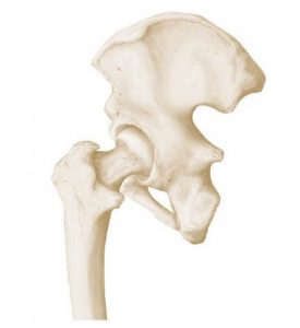
Anatomy: Hip Joint
The hip joint, otherwise known as pelvis or coxa, is a strong, large, weight-bearing joint. It is known as a ball-and-socket synovial joint, which means that it is composed of a bone with a rounded end (like a ball) and a bone with a carved out hole–these two ends fit perfectly together and allow for movement across all axes. The shoulder joint (glenohumeral joint) is also a ball-and-socket type joint.
The hip joint is made up of bone, cartilage, ligaments, tendons, muscles, blood vessels and nerves.
Bones
- Femur- Thigh bone (with its ball-like head) connects into the hip socket
- Illium- Large, flat, round bone on the top of the hip–these are the bones you can feel below your waist
- Ischium- Round bone behind and on the lower end of hip–these are the bones that you sit on
- Pubis- Pubis or pubic bone curves forward from the lower end of the illium bone and connects down and back to the ischium
Where these three bones (illium, ischium and pubis) connect a divot is formed known as the “acetabulum.” This is the socket where the femur bone connects, forming the ball-and-socket synovial joint. Around the femur head is a smooth cartilage-covered surface known as the labrum that allows for smooth movements in the acetabulum.
As one becomes an adult, the ilium, ischium and pubis bones fuse together creating the image of one bone, but it is technically these three bones fused together that make up the hip.
Other bones in the hip region include the sacrum and coccyx (tailbone).
Ligaments
Ligaments are the strong elastic structures that connect bone to bone providing strength and stability. In the hip joint there are three tough, strong ligaments that surround the joint capsule–encasing the ball-and-socket joint.
- Iliofemoral- connects the ilium to the femur
- Ischiofemoral- connects the ischium to the femur
- Pubofemoral- connects the pubis to the femur
Muscles
Muscles that surround the hip also help to provide strength and stability to this weight-bearing joint. There are actually no muscles that attach right into the hip joint–one of the reasons it is a stronger joint. The muscles that play important roles in the hip joint are in the lower back and thigh.
Nerves
- Sciatic Nerve
- Obturator Nerve
- Femoral Nerve
- Lateral Femoral Cutaneous Nerve
Bursae
A bursa is a small sac that is filled with synovial fluid. These small sacs (as many as 20 in the hip joint) help to provide less friction between bones, ligaments, and tendons for smooth, painless movements.
Hip Joint Problems
Typically the hip joint is very resistant to damage due to its structure, size and shape; however, a strong force to the hip can cause fracture or other injury, osteoporosis can cause weakness and fracture, and other trauma to muscles, tendons or ligament can cause displacement or other injury.
It is important to treat hip problems promptly as the hip joint can cause secondary issues (such as posture issues in the knees, ankles or spine) in other parts of the body if left untreated.
Types of hip joint problems may include:
Aplasia of the Acetabulum– a structural or functional issue of the acetabulum, can be common in newborns with restricted intrauterine growth or breech birth
Bursitis- inflammation or infection in the bursa
Coxa Valga– bone deformity where the neck of the femur and the body of the femur form an angle that is wider than normal
Coxa Vara– bone deformity where the neck of the femur and the body of the femur form an angle that is more acute than normal
Dislocation (Congenital, Trauma-induced, or other causes)- when a bone is displaced from its normal position
Fracture– a break or crack in a bone
Labral Tear– a rupture in the labrum
Legg-Perthes Disease– a bone formation disorder
Osteoarthritis– a degenerative disease of bone cartilage
Osteoporosis- a bone disease where new bone growth cannot keep up with the normal breakdown and regeneration of bone (bone breaks down with little to no re-growth)
