The anatomy of the lumbar spine is quite complex. Here we will attempt to provide a brief overview of lumbar spinal anatomy. The lumbar spine makes up the the lower end of the spinal column. It consists of 5 lumbar vertebra that are numbered 1 through 5 from top to bottom i.e. L1, L2, L3, L4, and L5. The L5 vertebra is connected to the top of the sacrum (named the S1 segment) through an intervertebral disc.
To review, the purpose of the spine, as a whole, is to support the body so that we can stand upright. Secondarily, it protects the spinal cord (which is the extension of the brain), and all of the nerves that branch from the spinal cord. At the level of the lumbar spine, the spinal cord has ended (typically at the L1-2 level in an adult), and at this level exists what is called the cauda equina (latin for horse’s tail), which is a fluid filled sack which houses the nerves that allow you to control your bowel and bladder as well as to move and feel your legs.
For more detailed descriptions of anatomical terminology and spinal cord anatomy, please reference:
Lumbar Spine Anatomy
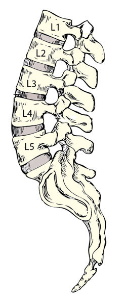
When looking from behind, in most individuals, the spine looks straight. However, when viewing the spine from the side, there are distinct curves to each part of the spine. The purpose of these curves is to grant some additional flexibility and shock absorbing abilities to the spine.
In the lumbar region, the spine normally curves backwards. This curvature is known as a lordosis. The amount of lordosis an individual has varies, but typically is somewhere in the range of 40-60 degrees. There are many conditions of the spine that may affect the normal curvature of the lumbar spine, resulting in pain and disability. Some of these conditions are listed at the end of this article.
Similar to the rest of the spine (thoracic and cervical), each of the vertebra of the lumbar spine consists of a body, two pedicles, lamina, and multiple bony projections (called processes). The vertebrae of the lumbar spine are the largest of the spine as they must support the most weight. The vertebral bodies are the major weight bearing portion of the vertebra. The processes serve as attachment points for various ligaments and muscles that are important to the stability of the spine.
The posterior (or back) aspect of the body, and medial (or inside) aspects of the pedicle, and the anterior (or front) lamina form a protective bony ring, called the spinal canal, around the very important dural sac. The dural sac contains all of the important nerves that allow you to control your bowel and bladder as well as to move and feel your legs. Each of these paired spinal nerves exits on both sides of the spine at each level of the lumbar spine. They exit between the pedicles (in an area called the intervertebral foramen). i.e. there are left and right L1, L2, etc. nerves that exit through intervertebral foramen at each respective level.
Between each vertebral body is a shock absorbing structure called an intervertebral disc. The intervertebral disc has two distinct components to it. The tough outer ring of the disc is called the annulus fibrosis. The soft, compressible inner portion of the disc is called the nucleus pulposus. The intervertebral disc is, in large part, is made up of water. This trait gives the disc much of its shock absorbing abilities. Unfortunately, as we age, the water content decreases, leading to degenerative disc disease, or more simply put, arthritis of the back. This is also a major reason why we become shorter as we age.
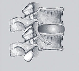
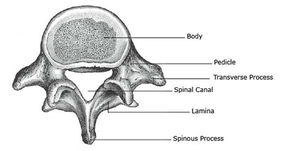
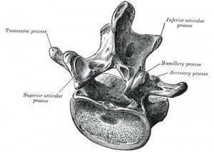
Behind the vertebral body are the various processes of the vertebra. These include the spinous process, the two transverse processes, and the paired superior and inferior articulating processes. As mentioned prior, each of these processes serve as attachment points for various muscles and ligaments which enhance the stability of the spine. Furthermore, the articulating processes of adjacent vertebra join to form facet joints, which, in the lumbar spine, allow for mainly bending and straightening of the spine.
Ligaments of the Back
As mentioned previously, the anatomy of the spine is quite complex. There are hundreds of individual ligaments in the spine. Ligaments are strong, tough bands that are typically not very flexible. Ligaments typically joint bones to other bones to stabilize them to one another. Some of these named ligaments in the spine include: the anterior and posterior longitudinal ligaments (ALL and PLL), interspinous ligaments, supraspinous ligaments, intertransverse ligaments, and ligamentum flavum.
Muscles of the Back
There are many muscles that help to both move and stabilize the spine. The major muscle group that allows us to stand upright is the erector spinae. This muscle group includes the iliocostalis, longissimus, and spinalis. Another important stabilizing muscle of the spine is the multifidus. There are many other smaller muscles of the spine and characterizing each one would be tedious and is beyond the scope of this article.
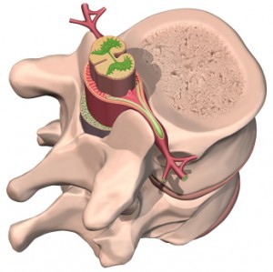
Vascular structures of the Back
The most important vessels of the spine include the anterior spinal and posterior spinal arteries. These vessels give blood supply to the nervous system within the spine. Additionally, each spinal unit has segmental vessels that come from the aorta that help to nourish the bony and soft tissue structures as well.
The Sacrum
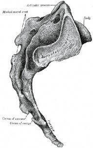
A discussion of the lower back wouldn’t be complete without an overview of the sacrum. At birth, the sacrum is actually made up of several vertebrae. By the time you’re an adult, these individual vertebrae have fused together to form the sacrum. By adulthood, it is a large, triangular bone, that forms the base of the spinal column where it connects to the pelvic bones. The top segment of the sacrum, labeled the S1 segment, connects to the L5 vertebra of the lumbar spine via the L5-S1 intervertebral disc.
Conditions of the Lumbar Spine
• Osteoporosis
• Osteomalacia
• Arthritis
• Ankylosing Spondylitis
• Sacroiliitis
• Lumbosacral sprain and strain
• Acute Cauda Equina Syndrome
• Intervertebral Disk Disease
• Scoliosis
• Spinal Instability
• Herniated Disk
• Spondylolysis
• Spondylolisthesis
• Spinal Stenosis
