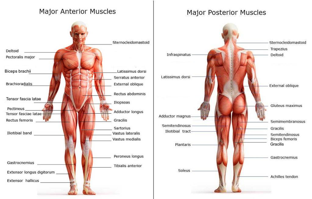
Our bodies have several groups of muscles. The chart below shows the outermost layer of those on the posterior side of the body. Some muscles function in opposition, both contracting (flexing) and straightening (extending). Others work as a group to perform the same function. The posterior muscles of the trunk support arm and back movements. They also protect the spinal column.
Muscles are often named based on their functions such as extensor, flexor, adductor, abductor. Tendons attach muscles to the nearby bones. They are elastic “band-like” structures that facilitate muscle movements.
Chart of Major Posterior Muscles

Achilles tendon
The Achilles is one of the strongest tendons in the body. It can support four times a person’s body weight. It is attached to the calcaneus (heel) bone, and is pulled by three flexor muscles: the soleus, the plantaris, and the gastrocnemius. This tendon is important for walking, running, and climbing stairs.
Adductor Magnus
This group of muscles is located on inner, posterior thigh, extending between the pelvic bone and the femur. It includes the hamstring muscles. Adductor magnus muscles provide stability while walking, and crossing of one leg over the other.
Biceps femoris
This muscle attaches and extends directly along the femur (thigh bone). It is one of the hamstring muscles, and is commonly injured while playing sports. The biceps femoris works in conjunction with the semitendinous and semimembranous muscles.
Carpi flexor ulnaris
This is a forearm muscle that is important for hand movements. Its tendon attachments are unique. On the upper arm, the tendon splits to attach onto two areas of the humerus. The distal end of the muscle attaches to two wrist bones and the fifth metacarpal.
Deltoid
Deltoid means a “triangular” shape. This powerful muscle wraps around the shoulder joint, and connects three bones: the scapula, clavicle, and humerus. Most shoulder and upper arm movements involve the deltoid muscle. It steadies the shoulder, and enables 360 degree arm movements:
- anterior fibers – arm flexion
- lateral fibers – arm abduction
- posterior fibers – arm extension
External oblique
This is the outer portion the abdominal muscles that are responsible for stability of the trunk. It also assists with twisting at the waist.
Gastrocnemius
The gastrocnemius is a flexor muscle that bends the foot downward such as when a dancer points her toes. It is more frequently known as the “calf” muscle as its name translates as “belly of the leg.” This muscle has two upper halves which attach to each side of the lower femur. Its distal end is the Achilles tendon which attaches the heel. As this muscle contracts, the foot points downward. It also supports bending at the knee.
Gluteus maximus
This outermost muscle of the buttocks is one of the largest in the body. It extends from the hip bones to the sacrum and coccyx. The gluteus maximus works in conjunction with the gluteus medius and minimus muscles to enable walking, standing, running, and climbing stairs. It also assists with posture by supporting the pelvis and trunk.
Gracilis
The gracilis is a very thin muscle that extends from the pelvis to the tibia in the lower leg. It is important for both hip and knee movements. When “groin” sports injuries occur, this muscle is most commonly affected.
Iliotibial tract
This muscle is also known as the iliotibial band, and extends from the hip the the lateral side of the distal femur (thigh bone). It also interacts with the gluteus muscles to enable leg movements at the hip, and extension and flexion of the knee.
Infraspinatus
The infraspinatus is the triangular-shaped muscle of the external rotator cuff in the shoulder. It attaches to the lower portion of the scapula, and stabilizes the humerus within the shoulder joint. Its primary function is abduction and external rotation of the arm.
Latissimus dorsi
The latissimus dorsi is a large muscle band that lies along the flank areas of the body. It originates on the lower thoracic and lumbar vertebrae, and attaches to the scapula, humerus, and iliac crest. In addition to internal rotation and adduction of the arm, the latissimus dorsi can also assist with breathing. In situations of respiratory distress, this muscle may be used by default to improve air entry into the lungs.
Plantaris Soleus
This very thin muscle extends from the knee to the heel, and works in conjunction with the gastrocnemius muscle. It helps with knee flexion, and upward pointing of the foot.
Sacrospinalis
Each sacrospinalis muscle lies along the vertebral column. They are also known as the erector spinae muscles. They are responsible for posture, and all spinal movements.
Semimembranosus
As one of the hamstring muscles, it extends along the back of the thigh from the hip to the tibia. It assists with leg extension and knee rotation.
Semitendinosus
This third hamstring muscle lies next to the biceps femoris. It works in conjunction with the semimembranous muscle to extend the leg, and flex at the knee.
Sternocleidomastoid
The SCM is the largest of the 20 neck muscles. There is one on each side of the neck which extends from the posterior lower jaw to the sternum. Working together, they support the head, and allow it to turn from side to side, flex forward, and extend backward. They also stabilize the temperomandibular joint in order to chew food properly.
Tensor fasciae latae
This lateral thigh muscle assists the gluteus muscle group and the iliotibial tract to stabilize hip and knee movements.
Teres minor
The teres minor is another of the rotator cuff muscles. It lies just lateral to the infraspinatous muscle, and assists with arm rotation. It also helps to stabilize the shoulder and prevent dislocations.
Trapezius
This large muscle group covers the upper portion of the back as well as the posterior shoulders and neck. When it contracts, the head tilts back, and the shoulders lift up. Each trapezius muscle extends from the lower thoracic spine to the base of the skull, and across the back and shoulders. Together, they form a four-sided, trapezoid shape, hence the name.
Triceps Brachii
The triceps brachia is the muscle that extends down the back of the upper arm. It straightens the forearm, opposing the contractions of the biceps muscle. This muscle is unique in that its proximal tendon divides into three smaller ones that originate on the scapula and two areas of the humerus. The lower end of this muscle is attached to the ulna by a tendon that allows elbow movements.
Glossary
Adducting
Movement that is toward the body. For example, the process of bringing the right leg next to the left one after it has been stretched to the side of the body. An easy way to remember this is that adducting “adds” to the body.
Extending
The act of straightening a limb, or reversing a flexed position. Extending the knee involves straightening the lower leg.
Inserts
Attaches to the bone.
Innervated
Supplied by a nerve.
Internal rotation
Rotating the anterior surface of a bone or joint toward the midline of the body. This is also called medial rotation. For example, internal rotation of the leg involves turning it so that the knee and foot point inward.
Originates
The proximal attachment to a bone.