The nervous system is divided into two systems:
The Central Nervous System (CNS) consists of the brain and the spinal cord. The spinal cord is the continuation of the brain which lies protected within the bones of the spine. You can think of the CNS as the control center for the body. It allows us to think, create memories, speak, move, run, etc.
The Peripheral Nervous System (PNS) consists of 12 cranial nerves, and 31 pairs of spinal nerves. The PNS acts as the system of electrical wires that allows for communication between the CNS and the body’s muscles and sensory receptors. They also control the automatic functions of the bowel, bladder, respiratory (breathing), and heart function.
Essentially, the PNS sends signals from the brain to the muscles of our body so that they can contract, which leads to movement. The PNS also relays information regarding sensation from the sensory receptors of our body to our brain, so we can feel and interact with the environment around us.
The spinal nerves are named according to where in the spine they emerge i.e. they are named in accordance with the level of the spine they exit from. E.g. the C2 nerve exits between the C1-2 vertebrae, the L4 nerve exits between the L4-5 vertebrae. As you can see in the 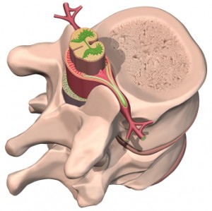 image to the left, the nerve roots (in red) branch off from the spinal cord to create the electrical wires of the PNS.
It will be helpful to review the anatomy of the cervical and lumbar spines before continuing on below.
Spinal Nerves
There are 31 pairs of spinal nerves. Again, they are named according to where they each exit in the spine (see figure below).

Each spinal nerve is attached to the spinal cord by two roots: a dorsal (or posterior) root which relays sensory information and a ventral (or anterior) root which relays motor information. Therefore, once the two roots come together to form the spinal nerve, the nerve carries a combination of both sensory and motor information (i.e. it contains mixed fibers).
The fibers of the sensory root carry sensory impulses from the body to the spinal cord, which ultimately relays that information to the brain. Sensory impulses include — pain, temperature, vibration, touch, and position sense (proprioception)—from organs, tendons, joints, and body surfaces.
There is a specific pattern to how nerves carry sensory information from our skin to our brain. Each spinal nerve carries sensory information from specific regions of our skin. These regions are called dermatomes (see below)

The motor roots carry impulses from the brain and spinal cord to the muscles of the body. These allow us to control the many muscles in our bodies.
The spinal nerves are divided into four main categories of spinal nerves based on the location from which they branch
- 8 cervical (C1-C8) nerves emerge from the cervical spine (neck)
- 12 thoracic (T1-T12) nerves emerge from the thoracic spine (mid back)
- 5 lumbar (L1-L5) nerves emerge from the lumbar spine (lower back)
- 5 sacral (S1-S5) nerves emerge from the sacrum (the triangular bone at the base of the spine)
- 1 coccygeal nerve emerges from the coccyx (the tailbone)
Below is a chart that outlines the main functions of each of the spine nerve roots:
| Spinal Nerve Root | Main Muscles Innervated | Other Notable Functions |
| C1 | Rectus capitis anterior / lateralis | |
| C2 | Longus capitis / longus cervicis / scalene | |
| C3 | Levator scapulae, rhomboids | Diaphragm |
| C4 | Levator scapulae, rhomboids | Diaphragm |
| C5 | Levator scapulae, rhomboids, deltoids, rotator cuff muscles | Diaphragm, biceps reflex |
| C6 | Biceps, wrist extensors (e.g. extensor carpi radialis brevis & longus) | Brachioradialis reflex |
| C7 | Triceps, wrist flexors (e.g. flexor carpi radialis, flexor digitorum superficialis) | Triceps reflex |
| C8 | Finger extensors (e.g. extensor pollicis longus) | |
| T1 | Finger abductors / adductors (e.g. interossei, lumbricals) | Sympathetic nerve output to the viscera |
| T2-T12 | Sympathetic nerve output to the viscera | |
| L1 | Sympathetic nerve output to the viscera | |
| L2 | Hip flexors | |
| L3 | Hip flexors, quadriceps | |
| L4 | Quadriceps, anterior tibialis, gluteal muscles | Quadriceps reflex |
| L5 | Extensor hallucis longus, hip abductors, gluteal muscles | |
| S1 | Gastrocsoleus complex | Achilles reflex |
| S2-4 | Parasympathetic nerve output to viscera | |
Nerve Plexuses
It is important to mention that after the spinal nerves exit from the spine, they join together to form four paired clusters of nerves, or nerve networks, called plexuses.
- Cervical Plexus – the cervical plexus represents the continuation of the upper cervical spinal nerves that gives innervation (i.e. supplies nerve function) to the neck and shoulders
- Brachial Plexus – the brachial plexus represents the continuation of the lower cervical spinal nerves that gives innervation to the upper back, shoulder, arms, and hand.
- Lumbar Plexus – the lumbar plexus represents the continuation of lumbar spinal nerves that give innervation to the lower extremities
- Sacral Plexus – the sacral plexus gives innervation to the back of the thigh, leg, bottom of the foot, as well as the pelvis