The following information will explain some of the causes of low back pain, how it is diagnosed, and describe the treatment options available.
Introduction
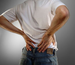 Back pain is the number one problem facing the workforce in the United States today. It is estimated that over 80 billion dollars is spent on back pain each year– and the cost is growing. Eighty percent of the population will suffer back pain some time in their life. These episodes usually occur between the ages of 30 to 50, the most productive period of life for most people. Most episodes of low back pain will resolve regardless of the type of treatment. However, a significant number of these episodes become chronic, meaning that they do not resolve. They continue to cause pain and dysfunction indefinitely. Back pain is a common chronic problem or many people, no matter what their occupation. There is no quick fix or total cure for most back problems.
Over many years your back is subjected to repeated stress which may not cause you pain at the time of the injury. However, repeated injuries add up, and eventually result in degeneration of the spine that causes lower back pain. Most episodes of low back pain are partially the result of degenerative changes that occur in the back. There may be an acute injury associated with the each episode of back pain. The overall condition of the lumbar spine usually determines how fast you recover from an injury and if your back pain will become a chronic problem.
The goal of a treatment program is to improve the current problem and to slow down the progress of the degenerative process. The physician’s role in the treatment of low back pain should be aimed at identifying the main problems that need immediate attention. He will also attempt to prevent your pain from becoming chronic by teacing you how to prevent further injury to your back. The purpose of this information is to help you understand more about your back problem so that you can make the decisions that will best help you to prevent injury or speed healing.
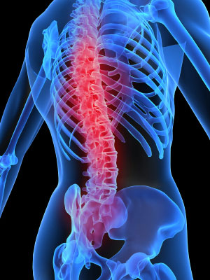
Anatomy – The parts of your spine and how they work
The Lumbar Spine is the last five vertebra of the spine. The vertebrae are the bones that make up the spine. They provide support and protection to the spinal cord. A joint is where two or more bones “join” together. Facet joints link the vertebrae together like a chain. They provide mobile connections between each vertebra. An intervertebral disk sits between each vertebra. The disk is actually a large, round ligament that connects the vertebrae to each other. If we look at a cross of the intervertebral disk, we can see that it is made of two parts. The annulus is the outer ring. It is the strongest part of the disk. It is responsible for connecting the vertebrae. The nucleus pulposus is the soft, inner portion of the disk. The nucleus has a consistency similar to crabmeat. The nucleus acts a as a shock absorber for the spine. The nerve roots of the spine carry information back and forth between the brain and the legs. It is pressure on these nerve roots that can cause pain and numbness.
 To better understand how the parts of the spine work together we will take a closer look at a spinal segment. A spinal segment is made of two vertebrae, the intervertebral disk between the vertebrae, and the two nerve roots, one from each side, exit from the spine. Notice how the facet joints fit together at the spinal segment. Looking at a cross section may be helpful.
Many of the problems that cause back pain result from an injury and the degeneration of the intervertebral disk. Degeneration is a wear and tear process that causes the disk to wear out, like when your favorite jeans get old. As we use our backs each day, the disk is put through many kinds of stress. The disk responds to the stress from the vertebrae by acting as a shock absorber. When we bend over, the disk is compressed by the vertebrae. This compression causes the disk to bulge outward toward the spinal canal and the nerves roots.
The facet joints also shift to allow the back to bend. And, like other joints in the body, the facet joints can be affected by arthritis.
Arthritis
The first changes that occur in the disk are tears in the annulus. The tears in the disk heal by forming scar tissue. Scar tissue is weaker than normal tissue. Repeated injuries to the disk cause the disk start wearing out. As the disk wears away, it loses some of its water content. It becomes stiff and no longer able to act as a shock absorber. If the disk continues to wear it will collapse causing bone to rub against bone. Bone spurs form as the disk wears out. We can watch the wearing out process by looking at a cross section of the spinal segment.
The nucleus of the disk loses water content and begins to break down. Then bone spurs form around the disk and the facet joints. These spurs are thought to be caused by the excess motion at the spinal segment. Eventually, bone spurs form around the nerves of the spine as well.
Herniated Disk
One of the most dramatic injuries to the lumbar spine is a herniated disk . In this injury, a tear in the annular ligament allows the nucleus pulposus to squeeze into the spinal canal. If there is pressure on the nerve root from the herniated disk, there is pain, numbness, and weakness along the nerve route.
Segmental Instability
The shift of one vertebra on another, called segmental instability can pinch the nerve root where it passes through the foramen. The excess motion can irritate the facet joints and cause mechanical pain from arthritis. Finally, the degenerating disk itself can become inflamed and cause mechanical pain.
Spinal Stenosis
In the late stages of spinal degeneration, bony spurs from the degenerative process can cause a condition known as spinal stenosis. As the bone spurs form, the size of the spinal canal becomes tighter around the spinal cord, pressing on the nerve roots. The pressure on the nerves cause pain and numbness in the legs.
Symptoms – How you perceive spine problems
Let’s use what you have learned about the anatomy to help you understand the symptoms of low back pain. Low back pain can be divided into two main types:
- Mechanical pain
- Compressive pain
Mechanical back pain results from movement of the back. Mechanical back pain is comes from inflammation caused by irritation or injury to the disk, facet joints, ligaments, or muscles of the back. A common cause of mechanical pain is disk degeneration. Mechanical pain usually starts near the lumbar spine. Mechanical pain can also spread to include the buttock and thigh areas. However, it rarely goes below the knee.
Compressive pain occurs when nerve roots that leave the spine are irritated or pinched. A common cause of compressive pain is a herniated disk. The nerves that leave the lower lumbar spine join to form the sciatic nerve. The sciatic nerve provides sensation and controls the muscles of the lower leg. Pressure on the nerve roots of the lumbar spine can interfere with the normal function of the sciatic nerve. An early sign of pressure on a nerve root is numbness along the
route of the nerve. There is also pain in the same area, usually extending below the knee to the foot. It is not unusual for the back itself to be pain free. Finally, the muscles that the nerve controls can become weak and the reflexes disappear.
Also, spinal stenosis can cause compressive pain. In some people, degeneration of the spine results in a narrowing of the spinal canal near the spinal nerves. This causes all of the nerves inside the spinal canal to become inflamed, and not function as they should. You may feel numbness in one or both of your legs below the knee. The numbness may get worse with activity such as walking. Also, pain can involve both of your lower legs. The pain gets worse with activity and gets better with rest. Weakness of the muscles of both legs may also occur. Again, this weakness can get worse with an increase in activity.
The Cauda Equina Syndrome
WARNING!
In a very few cases, a disk herniation can be so severe that it fills the entire spinal canal. A sudden pressure on the nerves at that level may cause a loss of control of the muscles that involuntarily control the bowels and bladder. If you lose control over your bowels or bladder, you should contact your physician immediately.
THIS IS AN EMERGENCY!
Diagnosis – How we look into your back problem
During your first visit, your doctor ask you to describe the complete history of your back pain. Then he will give you a thorough physical exam. There are many tests your doctor can use to find out the cause your low back pain. He will probably order x-rays of your lower back. X-rays are a first step in looking into any back problem and will help decide if more tests will be needed.
X-rays show the bones of the lumbar spine. Most of the soft tissue structures of the spine, such as the nerves, disk, and muscles, do not show up on x-ray. Still, much can be learned from the x-rays. Here we see X-rays taken of the same patient 20 years apart, showing how much the spinal segments have degenerated during that period of time.
Magnetic Resonance Imaging (MRI)
The MRI (magnetic resonance imaging) scan is the most common test used to evaluate the spine. The MRI is a scanner that uses magnetic waves to see the
structures of the back. Imagine that you could cut the spine into many small slices and then take a picture of each slice. The MRI scanner allows the doctor to take lots of “picture slices” of the spine without actually cutting the spine. The MRI is better than xray because it allows the doctor to see the bones and the nerves and disks; the nerves and disk could not be seen on an xray.
Picture slices can also be taken across the spine, giving a cross section view. The MRI scanner allows us to clearly see the nerves and disk without special dyes or needles. Here we see cross section MRI that shows a side view and cross section view of a common type of herniated disk.
Computer Assisted Tomography (CAT Scan)
The CAT Scan is an x-ray very similar to the MRI Scan. Instead of picture slices, x-ray slices can be taken across the spine, giving a cross section view of the spine. The CAT Scan shows the bones of the spine much better than the MRI. The CAT scan is useful when conditions that only affect the bones of the spine are suspected. The CAT Scan is commonly combined with a Myelogram to give a better picture of the spinal nerves and help determine if the is pressure is being caused by spinal stenosis or a herniated disk.
Myelogram
A Myelogram is an x-ray where a special die, which can be seen on the xray, is placed into the spinal sac. If the shape of the spinal sac looks abnormal, or indented, this may mean there is pressure on the nerves of the spine. A herniated disk may cause this pressure.
Discogram
The Discogram is a special test where dye is injected directly into the nucleus pulposus of the disk. The injection may cause you to have the same type of low back pain you have been having. If so, the pain suggests that the disk being tested is the one causing the pain. Once your doctor finds the disk causing the pain, regular xrays and a CAT Scan can be used to see if the disk has ruptured.
Electromyogram
An Electromyogram (EMG) is a test that looks at the function of the nerve roots leaving the spine. The test is done by inserting tiny electrodes into the muscles of the lower leg. The EMG can detect abnormal electrical signals in the muscles. The EMG can show if a nerve is being pinched after it branches off the spine.
You can compare the EMG to how you would test the wiring of a lamp. If you place a good light bulb into the lamp, and the bulb lights up, you assume that the wiring is OK. But if the bulb doesn’t light up you can safely assume that something is probably wrong with the wiring. The lamp may be unplugged or a short circuit may have occurred. The leg muscles are like the light bulb in the lamp. The EMG is can tell the condition of the nerves that supply those muscles, much like the wiring on the lamp. If the EMG finds that the muscles (the light bulb) are not working as they should, we can assume that the nerves (the wiring) are getting pinched somewhere.
Bone Scan
A Bone Scan helps find the area of the spine that is not normal. To do a bone scan, a radioactive chemical is injected into the bloodstream. This radioactive chemical attaches itself to bones that are undergoing rapid changes. The areas of the skeleton that are undergoing rapid changes appear as dark areas. Once the affected area is identified, other tests, such as the MRI are done to look closer at the specific area.
There are many possible causes of low back pain; some are not related to degeneration of the spine. Blood tests to look for infection, or arthritis may be necessary. Problems from areas other than the spine can cause back pain. These can include: aortic aneurysm, kidney problems, and stomach ulcers. Your doctor may order specific tests to find out if the cause of your back pain is from something other than your back.
Treatment – Options available to help you with your back problem.
The treatment of back pain can range from the reassurance that nothing is wrong to very delicate surgery. No two people are the same. Treatment must be based on the individual and their symptoms. In general, treatment falls into two major categories:
- Conservative treatment —which includes exercise, medications, physical therapy, and other non-surgical treatment.
- Surgical treatment—which includes laminectomy, diskectomy, and spinal fusion.
Surgery is only necessary for a few patients. There is no single surgery that will work for all spine problems. If your doctor thinks surgery ill improve your problem, he will suggest the type of surgery he thinks is the best for your specific problem.
Treatment for any back condition should involve two goals:
- Pain relief, and
- To reduce the risk of re-injury
Conservative Treatment
Exercise plays an important role in achieving pain relief and reducing the risk of injury. Many studies have shown that people who exercise regularly have far fewer problems with back pain. Exercise stimulates the body’s natural pain controlling hormones and actually decreases the perception of pain. Your doctor may have a Physical Therapist help you with an exercise program. The physical therapist will teach you ways to prevent further injury to your back.
Medicine should be used wisely!
- Some medicines are highly addictive!
- No pain medicine will control chronic pain if used over a long period of time
- No medicine will cure back pain caused by degenerative origin.
- Medications are used to control: pain, inflammation, muscle spasm, and sleep disturbance.
If simple measures fail to relieve your back pain you may be given an Epidural Steroid Injection (ESI). There are many different causes for inflammation of the nerves in the lumbar spine. An epidural steroid is an injection that puts a small amount of cortisone into the bony spinal canal. Cortisone is a very strong anti-inflammatory medicine that may control the inflammation surrounding the nerves and ease your pain. The epidural steroid injection is not always successful and works about 40% to 50% of the time. These injections are only used when all other conservative measures have failed, or as a last attempt to postpone back surgery.
Surgical Treatment
Diskectomy
One of the most common surgical procedures is a diskectomy. A diskectomy relieves pressure on a nerve root removes the herniated disk that is causing the pressure. First, a portion of the lamina of the vertebra is removed. Remember, the lamina is the back part of the vertebra and forms a roof over the the spinal nerve. Removing a section of the lamina leaves a “window” into the spine. The spinal nerves are then moved aside so that the herniated disk can be seen. Then small instruments are used to remove most of the nucleus pulposus to keep the disk from herniating again. Once the disk material is removed, the nerves are free from pressure and irritation. Both the lamina and the part of the disk that was removed will fill with scar tissue very quickly.
Laminectomy
If spinal stenosis seems to be the major cause of your pain, then spinal canal needs to be made larger. This is usually done by performing a complete laminectomy. A laminectomy removes all of the lamina. It may be easier to understand by looking at a cross section. Removing the lamina allows more room for the nerves, and also allows the surgeon to remove bone spurs from around the nerves. This allows more room for the nerves of the spine and reduces the irritation and inflammation of the spinal nerves. The lamina does not grow back. Instead, scar tissue grows over the bone, replaces the lamina, and protects the spinal nerves.
Spinal Fusion
If your back problem is the result of segmental instability, a spinal fusion may be recommended. A spinal fusion is done by placing a bone graft between two or more vertebrae, causing the vertebrae to grow together, or fuse. The bone graft is usually taken from the bones of the pelvis at the time of surgery. There are two types of spinal fusion:
- Posterior fusion – In the posterior fusion, the bone graft is placed on the back side of the vertebrae. During the healing process the vertebrae grow together creating a solid piece of bone between the vertebrae.
- Interbody fusion – The interbody fusion differs by replacing the removed disk with a bone graft between the vertebrae.
Once again, the healing process causes the vertebrae to grow together creating a solid piece of bone between the fused vertebrae.
In both types of fusion the vertebrae grow together into one bone. The goal of a spinal fusion is to stop the motion from segmental instability. This reduces the mechanical back pain and impingement on the nerve root caused from excess motion.