Evolution and Types of Eye Surgery
Corrective surgical procedures for human eye have been practicing for more than 100 years. However, techniques for corrective eye procedures are improving from last few decades –especially after the discovery of laser procedures. Artificial lenses are used to correct the refractive errors of the eye. With the help of refractive eye surgery, the refractive errors are corrected either by modifying the shape of the cornea or a man-made lens is inserted into the eye. Thus, refractive surgery helps to eliminate or reduce the use of extraocular lenses. Refractive errors such as myopia, hyperopia, and astigmatism are treated with the help of surgical procedures.

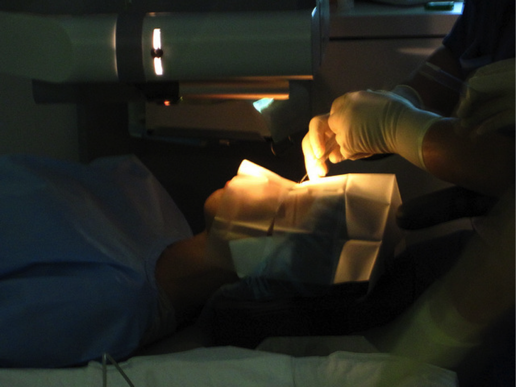
Eye Surgery Image courtesy: flickr.com
Radial Keratotomy
In 1898, keratotomy was explained by L.J. Lans from Netherlands. The procedure was further studied during 1940s in a clinical environment by K. Akivam and T. Sako of Japan. In 1960s, Slava Fyodorov of USSR increased the safety of the keratotomy procedure by creating multiple incisions on the cornea to form a clear optical zone in the center. In 1978, Radial Keratotomy was introduced in the United States. Radial Keratotomy was used to rectify myopia (nearsightedness). In this procedure, radial incisions are created on the outer surface of the cornea to change the shape of the cornea. The radial incisions made using steel surgical blades flatten the cornea in the center. The procedure changes the focal length of the cornea and thereby corrects the refractive error. In some people, radial keratotomy created problems such as fluctuating vision, glare, regression, and night vision problems. These side effects were more prominent in patients with higher prescription, whereas the side effects were rare in lower prescription patients. The major disadvantage of radial keratotomy is the excessive time required for recovery.
Automated Lamellar Keratoplasty
ALK was mainly performed in hyperopia (farsightedness) patients and also in patients with severe myopic condition. Automated lamellar keratoplasty is a surgical procedure introduced during mid-1990s in which a microkeratome is used to cut the corneal flap. The corneal flap then folded to one side and a thin slice of corneal surface tissue is removed. The removal of corneal tissue changes the refraction as the procedure alters the shape of the cornea. Some of the risks involved in this procedure are overcorrection, astigmatism, under correction, scarring, loss of corneal flap, loss of vision, infection, and glare.
Photorefractive Keratectomy
Photorefractive keratectomy or PRK is the first successful laser procedure. This surgical procedure is used to change the corneal curvature using laser directly on the cornea tissue to remove the surface tissue. PRK is used to correct low to moderate hyperopia, low to high myopia, and astigmatism. Even though PRK was introduced in the United States in 1980s, it got approval from FDA in the year 1995. This procedure is widely used even today, but the most popular surgical procedure is LASIK. PRK does not pose any risk of corneal flap complications as the procedure is not performed by creating a hinged corneal flap. PRK is safe for people with thin cornea. Since the laser is used directly on the surface tissue of the cornea, the recovery time for the procedure is lengthy.
LASEK
LASEK or Laser Assisted in-situ Epithelial Keratomolysis is invented in the year 1999 by an Italian ophthalmologist, Massimo Camelin. This procedure fixes the refractive errors such as myopia, hyperopia, and astigmatism. In this procedure, the epithelium is peeled back and the cornea is exposed. After reshaping the cornea using the excimer laser, the epithelial layer is placed back. The recovery time of the procedure is fast, but the discomfort is more when compared to the LASIK procedure.
LASIK or Laser Assisted in-situ Keratomileusis is introduced in 1989 by a Greek doctor, Ioannis Pallikaris. However, the FDA gave approval to the procedure only in 1999. In LASIK procedure, laser is used to create the corneal flap and then corrects the curvature of the cornea. Recovery is very quick and there will be no discomfort after the procedure.
Phakic Intraocular Lens – Visian ICL
Phakic intraocular lens is inserted inside the eye without any tissue removal. It works along with the natural lens. Even though this artificial lens insertion is a permanent solution, it can be removed if required. The lens was developed by Fyodorov Institute, Russia in the year 1992.