Anatomy and Function of the Knee
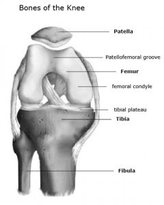
Before you start, it will be very helpful to review our article on knee anatomy to better understand the anatomy of the knee, where the patella (knee cap) is located, and how the knee functions.
Anatomy and Function of the Patella
The patella, or kneecap, is the circular and more mobile bone that exists on the front of the knee. The patella is part of the extensor mechanism of the leg; a major stabilizer of the knee and the primary extensor of the knee (i.e. straightens the knee). The extensor mechanism is composed of the quadriceps, the quadriceps tendon, patella and patellar tendon, and their associated soft tissues. The patella gives mechanical advantage to the quadriceps muscle and enables it to more powerfully straighten out the knee.
In order for the patella to track up and down normally within the patellofemoral groove (formed by the medial and lateral condyles of the femur), there has to be a balanced pull from both the lateral (outside) and medial (inside) sides of the patella. This balance is achieved by the overall alignment of the leg and the associated soft tissues around the knee. Importantly, a patella that stays centered within the patellofemoral groove throughout the knee’s full range of motion is a well-functioning patella.
The underside of the patella is covered with the thickest amount of articular cartilage found anywhere in the body. Articular cartilage is a smooth and spongy covering that is found on almost all joint surfaces. This slippery surface helps the patella glide within the patellofemoral groove of the femur as the knee goes through motion.
How The Patella Causes Knee Pain
People can develop knee pain when the cartilage under the patella begins to wear and tear. This results in softening of the cartilage (called chondromalacia patellae). Chondromalacia can develop for numerous reasons. Age (generalized wear and tear — similar to putting a lot of mileage on a car), trauma, or muscle imbalances around the knee can all contribute to the development of chondromalacia.
A very common knee problem is when the patella moves slightly abnormally as the knee bends or straightens. This happens because of an underlying muscle or structural imbalance around the knee. Normally the patella moves centrally within the patellofemoral groove — a path controlled by the quadriceps muscle and the patellar tendon. Abnormal movement can cause wear and tear to the cartilage.
These aforementioned structural imbalances can exist if there are variations in the anatomy of the bones in the thigh or leg or due to increases in the Q-angle — the angle created between a line through the center of the quadriceps tendon and a line through the center of the patellar tendon. With an increased angle, the patella naturally wants to be pulled outwards out of the patellofemoral groove as the knee goes through range of motion. Women tend to have a greater angle than men.
Another structural variation that can contribute to knee issues is when the the lateral (outer) femoral condyle is smaller than normal. This forms the lateral “bunker” of the patello-femoral groove which keeps the patella tracking within the groove. If the femoral condyle is too small, this bunker may be non-existent. Consequently, the patella will have a tendency to slip out of the groove.
Abnormal mechanics of the patellofemoral joint can lead to altered pressures on the patellar cartilage which can lead to early chondromalacia and arthritis. It is also increases the risk of injury to the knee e.g. patellar dislocation.
Symptoms Of Patellar Problems
Depending on the underlying problem, symptoms vary. For instance, people with chondromalacia patellae may experience vague knee pain that is hard to localize. Pain is commonly associated with walking down stairs or hills, keeping the knee bent for long periods of time (i.e. prolonged sitting), or while kneeling. The knee may even feel as though it wants to gives out on occasion, but this is a reflex response to pain and not an indication of instability in the knee. Finally, you may feel that your knee grinds or pops when squatting or going up and down stairs. This occurs with motion as the normally smooth surfaces of the patella and femur become roughened and rub against one another.
Patellar instability can occur after a patellar dislocation. After the first dislocation, it may continue to want to dislocate, making it hard for you to trust your knee.
How Are Patella Problems Diagnosed
Diagnosis begins with a complete history i.e. your story regarding your knee pain, how it started, and what types of activities aggravate it. Your physician will examine you while standing, sitting, and/or lying down. Strength of the muscles around the knee will be tested. X-rays will likely be performed by your primary care doctor prior to visiting with an orthopedic surgeon. An x-ray helps doctors better understand the alignment of your knee and if there is any evidence of any bone issues like fractures or underlying arthritis.
If symptoms persist, however, a MRI scan may be needed to look more closely at the knee to see what might be causing your symptoms. Ultimately, if surgery is warranted, knee arthroscopy will additionally allow for the identification of any problems that were not seen on X-rays.
What Are The Treatments For Patella Problems
For the most common patellar problems, symptoms improve with a short period of rest, anti-inflammatory medication and physical therapy. Physical therapy typically consists of exercises to strengthen the muscles around the knee. Additionally, the therapists will work with you to improve upon your flexibility. Both of these factors will allow for a more well-balanced and a better functioning knee.
For instance, with chondromalacia patellae, usually the Vastus Medialis Obliqus (VMO) muscle is targeted for strengthening. While helps for improved mechanics of the patellofemoral joint.
If anti-inflammatory medications and physical therapy fail to address your issue. Depending on your underlying problem, your doctor may recommend a steroid injection. With a steroid injection, a strong anti-inflammatory is injected into the joint space.
If non-surgical treatments fail to improve your condition, surgery may be warranted. For instance, with patellar instability, the patella has a tendency to want to dislocate laterally (outwards). Usually this is due to a structural problem with your knee. A lateral release may be performed to improve the sliding mechanics of the patella — specifically, to ensure that it stays centered in the patellofemoral groove with motion. In a lateral release, the tight lateral ligaments on the outside of the patella are cut to allow the patella to move towards the center of the femoral groove.
Depending on the severity of your malalignment, the medial (inner) aspect of the patella may also need to be reinforced to further ensure that the patella stays centered with knee motion. This latter surgery is called a MPFL (medial patello-femoral ligament) reconstruction.
In very severe cases where the patella keeps dislocating, the attachment of the patellar tendon on the tibia may also have to be moved. Once the surgery heals, the patella will track centrally within the patellar groove, reducing pressure on the articular cartilage.

