Elbow X Ray: Definition
X ray is an imaging technique that uses small amounts of electromagnetic radiological waves, called “x” waves, to capture images of internal body structures like bone. An x ray image of the elbow joint provides information for assessment, evaluation and diagnosis of elbow joint disease process or injury, such as fracture and gives information key in developing a proper treatment plan. The elbow joint is defined as the point of connection or articulation between the humerus bone (upper arm) and the radius and ulna bones (forearm bones).
Elbow X Ray: Limitations
Typically an x ray is an effective imaging technique to assess, evaluate and confirm diagnosis of a variety of bone injury and damage in the elbow. However, if the problem is related to soft tissue structures such as muscle, ligament or tendon, another type of imaging may be prefered. In addition, if a more detailed diagnosis of infection or cancer in the bone is needed a bone scan may be recommended.
X Ray, MRI, Bone Scan or Ultrasound
The following is a brief description of some other imaging techniques that may be used in assessment, evaluation and diagnosis of bone pathology or disease when x ray may not seem appropriate.
Bone Scan
Bone scan is another imaging technique that is typically performed in the medical field known as nuclear medicine. It is used to show absorption patterns in bone in order to diagnose a variety of other diseases and disease progression. A small amount of a radioactive substance is injected into a vein and settles into bones. Depending on the absorption of this tracing substance, nuclear medicine specialists can diagnose abnormal metabolic activity in bones such as cancer, metastasis, fractures, infection and inflammation.
Magnetic Resonance Imaging
Magnetic Resonance Imaging (MRI) is another imaging technique used to capture detailed pictures inside of the human body. It uses magnet, radio waves and computer technology to take pictures of the body’s organs, bones or soft tissues, such as tendons, ligaments and muscles.
The main difference between MRI and x ray is that an MRI does not use radiation.
Ultrasound Imaging
An ultrasound uses ultrasound, high-frequency sound waves, to capture images of internal body organs, muscles, tendons, joints and blood vessels. A distinguishing feature of ultrasound imagine is that it can capture movement of internal structures in real time.
Who do I need to see to get an x ray?
Radiologists are specialized doctors that are educated in reading and interpreting findings of x ray images.
Often times a radiology technician will capture the desired and necessary images, helping to place the joint in the desired position, taking various images at different viewpoints and angles, and making sure the radiologist gets the images.
If you think you need an elbow x ray, speak with your primary care provider or seek medical attention at an urgent care center or emergency room. They will likely be able to help refer you to a radiologist, or take the images and have a radiologist read and interpret the findings to inform their diagnosis.
How is an x ray read or interpreted?
The x ray images are viewed by a specialist known as a radiologist. A radiologist must know what a normal elbow joint looks like in order to know what how an abnormal joint appears in an x ray image. Radiologists are educated and trained in detecting different types of fractures, blood flow patterns, bone measurements and densities, angle measurements, and foreign object detection among other findings and interpreting these findings to a physician.
Purpose of X-Ray
The purpose of an x ray may be to diagnose a disease or injury, or to assess, evaluate or monitor the progression of growth or disease.
Risks and Benefits of X-Ray Imaging
Risks of X-Ray
The risks of having an x ray image of the elbow joint are very minimal and low. Scientists and doctors over the years have improved the procedure greatly to use the smallest amount of radiation possible and have discovered ways of minimizing the radiation waves from scattering to other parts of the body.
In general the two main risk factors for x ray imaging are 1) development of cancer due to over exposure to radiation 2) harm to fetus and/or reproductive organs.
Cancer Risk
With respect to the risk of cancer due to x ray imaging: the risk increases with repeated exposure to x rays, or exposure to large amounts of x rays. In general, a few x ray images of an elbow joint is not enough to cause cancer.
Pregnancy Risk

With respect to the risk to pregnancy due to x ray imaging: always inform your doctor, the radiologist technician, and other members of your health care team if you are pregnant or planning to become pregnant before having an x ray. The risk increases with repeated exposure to x rays, or exposure to large amounts of x rays–especially to the abdomen.
Likely, if it is a localized area away from the abdomen that they are needing to x ray (like the elbow), you can still have an x ray image taken. You will be asked to wear a lead apron to protect your abdomen and reproductive organs from exposure of scattered x rays.
Benefits of Elbow X-Ray Imaging
X ray imaging is a quick, painless, and relatively inexpensive way to provide an assessment, evaluation and diagnosis of elbow pain, and/or immobility. When a bone fracture or dislocation is suspected an x ray is a quick way of helping to confirm a diagnosis.
Elbow X Ray: Positioning and Viewing
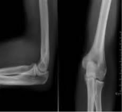
Typically images of the elbow joint are a combination of two positions: anterior-posterior (front-back) and lateral (side view).
Depending on the suspected issue or injury in the elbow joint, the radiologist may want to see the picture of a certain angle or position of the elbow.
Typically the images are either taken from front to back or back to front in a view known as anterior-posterior or AP. Or, the images are taken from a side view, known as lateral view.
The radiologist may want to see the elbow joint straight (extended) or bent (flexed) position, or a combination of angles depending on what they are looking for.
Elbow X-Ray: Cost
The cost of an elbow x ray will depend on your location, provider, how many images are taken and what type of insurance you have. However, an estimate cost of an elbow x ray can range from $100 or less to up to $1,000 (without insurance). A more exact estimate may be between $260-$460.
What can you see in an x ray?
Typically x ray viewing shows bone. In viewing a joint or bone through x ray imaging, one is able to make many observations (including angle measurements, length and width measurements, measurements of the cartilage space, the intactness of the bone, etcetera) to provide information for diagnosis and treatment.
When images of soft tissues (such as muscles, tendons, and ligaments) surrounding bone or bone joints is required, an MRI may be done instead.
Normal Findings
Anatomy: Elbow Joint/ Bones
The elbow joint is comprised of three main bones:
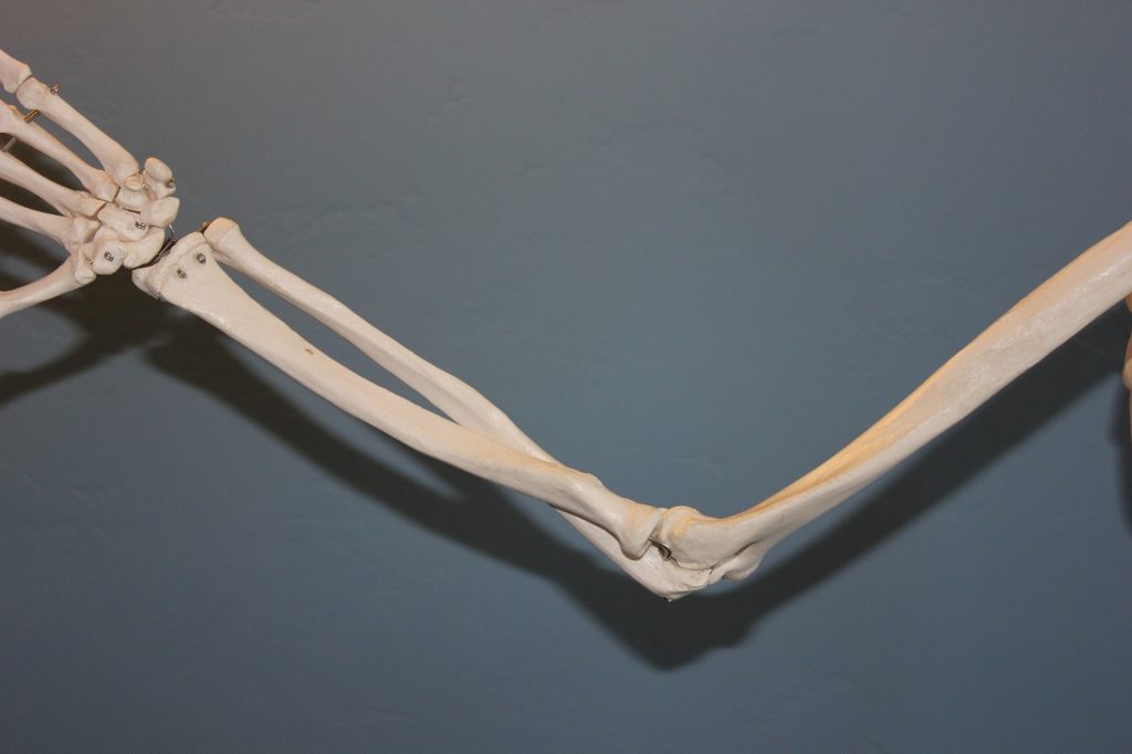
- Humerus–the long upper arm bone that connects the shoulder socket joint to radius-ulna joint.
- Radius–one of the forearm bones that connects at the humerus down to the thumb side of the hand. It allows for “radial” (circular, or twisting) movements.
- Ulna–one of the forearm bones that connects at the humerus down to the pinky side of the hand. The part of the elbow that is the main boney tip (commonly referred to as the “funny bone”), is the tip of the ulna. Medically it is known as the olecranon.
Abnormal Findings
What injuries might show up on an elbow x ray?
Types of arm bone/ elbow joint fractures
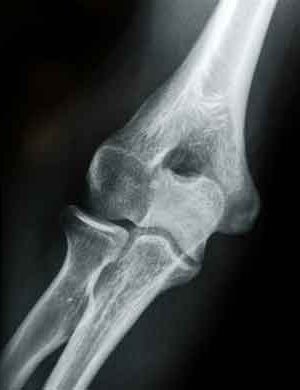
Olecranon fracture
An olecranon fracture refers to a break located in the olecranon, boney tip of the ulna bone. A less severe fracture may be treated by using a splint or cast for a period of time while the fracture heals itself. A more severe fracture, that often affects the placement of surrounding bones, requires surgery to initiate the repair process.
Symptoms of an olecranon fracture may include:
- Sudden, intense pain in the elbow
- Severe pain with movement (sometimes limiting ability to move joint)
- Numbness in one or more fingers
- Bruising and/or swelling around elbow tip
- Instability of the joint (feels as if the bone will ‘pop’ out of place)
Causes of an olecranon fracture:
Olecranon fractures can be fairly common. Usually this type of fracture is caused by falling and landing inappropriately or by direct forceful trauma to the elbow.
Elbow Fractures in Children
Elbow fractures or breaks can be common in children due to a higher rate of falling and engaging in activities that pose high risk of elbow injury (falling from monkey bars, sports falls, etcetera). It is important to remember, however that children may not be able to articulate that they fell or may have broken a bone in their elbow. If you note that a child is not moving their arm or cannot straighten their arm, assist them to seek medical attention promptly.
It is also important to note that when a child has sustained any form of injury, that it may be important to evaluate for the presence of physical abuse.
Different types of fractures and breaks are prevalent among children. These types of fractures are classified or named by their location.
Examples of elbow fractures in common in children include:
- Supracondylar- refers to a break or fracture of the humerus bone, just above the condyles (boney protrusions at the head of the bone)
- Condylar- refers to a break or fracture of the boney protrusions (knobs) at the end of the bone. There are condyles on the outer (lateral) and inner (medial) side of the humerus bone.
- Epicondylar- refers to a break or fracture of the inside of the elbow tip.
- Physis- refers to a break or fracture of the growth plate in either the upper arm or either forearm bone. Growth plates are areas of cartilage that serve to grow over time. Injury to this area, if not treated properly or quickly can cause disruption to proper growth can result in no growth and/or improper growth (deformity).
- Forearm- refers to a break or fracture of the radius or ulna bones. The head of the radius bone can break completely off or be susceptible to a compression fracture. The tip or head of the ulna bone is known as an olecranon fracture.
- Monteggia Fracture- refers to a break or fracture of the ulna (olecranon) with dislocation of the radius bone.
- Open Fracture- refers to a fracture or break of any bone that protrudes through or breaks the skin.
When injury, dislocation, fracture or break is suspected in the elbow joint it is very important to get it treated and evaluated quickly. Sometimes breaks affect more than just the injured bone and include injury to nerves, tendons, ligaments, or surrounding bones (dislocation). It is important to make sure that it is not just the broken bone that is treated, but all these other surrounding structures as well.
Types of Tendon Injury in Elbow
Tennis Elbow
In adults, one of the most common causes of elbow pain is related to a type of tendonitis (inflammation of the tendon) known as tennis elbow. Remember that tendons connect muscle to bone–in this case repetitive muscular movement of the bone causes strain and injury to the tendon. This injury is to the lateral (outer) tendon of the elbow.
Tennis elbow is caused by repetitive motions that include gripping. It is known as “tennis elbow” because it is common among tennis players to grip a racket and use repetitive swinging, twisting, hitting, bending motions with their elbow. It is not just tennis players, however, who are susceptible to tennis elbow. Any other job or activity that involves repetitive hand, wrist, and elbow movements such as knitting, typing, raking, painting or mopping can cause tendonitis around the elbow joint.
While tendon injury and inflammation may not show up on an elbow x ray, several x ray images may be captured in order to rule out other injury.
Golfer’s or Pitcher’s Elbow
Similarly to tennis elbow, golfer’s or pitcher’s elbow is another common tendon injury in the elbow joint. Known as medial epicondylitis, it affects the medial (inner) tendon of the elbow.
Types of Ligament Injury in Elbow
Ulnar Collateral Ligament Sprain or Tear
General elbow pain, stiffness or weakness may signify injury to the ulnar collateral ligament (ligament connecting the humerus to the ulna). Ulnar collateral ligament sprain is a common ligament injury in the elbow joint. Remember that ligaments connect bone to bone–breaks, dislocations and other injury can cause tearing (partial or full) of ligaments. Ligament injuries are also known as sprains.
Causes of injury to this ligament can include sudden twisting or tearing of the ligament due to a fall or blunt force; or due to stress on the ligament from overuse or repetitive use.
Pain, swelling and weakness can be symptoms of injury to the ligament. While this type of ligament injury may not show up on an elbow x ray, several x ray images may be captured in order to rule out other injury.
