Many people have the unfortunate experience of a head injury during in their lifetime. It can occur during a collision while playing sports, a fall on pavement or stairs, or a car accident. Both the immediate and subsequent symptoms are known as a traumatic brain injury (TBI). Mild injuries produce minor symptoms that may resolve within a few weeks. Moderate to severe head injuries can result in longterm health problems, and changes to the quality of life.
Brain Anatomy
The brain is our body’s “control center.” It is located inside of the skull, and is covered by a protective “skin” called the dura mater. Between the skull bones and this dura mater is cerebral spinal fluid. This fluid cushions the brain to protect it from injury, provides nutrients to brain cells, and removes cellular waste. In addition, blood vessels lie along the brain’s surface, and within the brain tissue itself. Any disruption of these blood vessels or layers of protection can impair brain function. Some injuries occur at the level of the brainstem. This is the narrow segment at the base of the brain that provides a connection to the spinal cord. Therefore, it is important to know which areas of the brain have been injured since each region controls different body functions.
Symptoms of a Brain Injury
A headache is the most common symptom. It may be associated with additional symptoms.based on the severity of the injury. Severity is often determined by whether or not there is a loss of consciousness at the time of injury.
- Mild Symptoms: nausea, dizziness, mood changes, and difficulty with focus, memory, and sleep
- Moderate Symptoms: severe headache, light sensitivity, vomiting, unsteadiness with walking
- Severe Symptoms: loss of consciousness, seizures, agitation, lethargy
In addition to these neurological symptoms, there may be obvious physical signs if injury such as bruising or swelling on the head.
How brain injuries are diagnosed.
A physical exam and diagnostic tests are used to determine the location and extent of damage to the brain.

These exams and tests can include some or all the following:
– A neurological exam is a series of questions and simple commands asked by a primary care doctor, trauma specialist, or neurologist to assess how well a patient can open the eyes, move, speak, and understand what has occurred. Examples include, “What is your name?” “Where are you?” “ What day is it?” “ Wiggle your toes,” or “Hold up two fingers.” The injured patient’s reflexes, muscle strength, and coordination are also evaluated. For example, the patient may be asked to walk in a straight line or stand with both eyes closed to identify problems with maintaining balance. The patient’s eyes will also be checked for normal pupil reaction, and for pressure on the optic nerve.
– Regular x-rays are images that reveal fractured or broken bones This modality is often used to look for skull fractures in infants because of the lower radiation exposure.
– A CT (CAT) Scan is a painless but stronger x-ray of the brain or other parts of the body. It provides very detailed images of the skull to locate fractures. It can also detect areas of bleeding, pressure on the brain tissue, and other abnormalities. CT scans emit a greater amount of radiation than x-rays, so their use is reserved for moderate to severe head injuries.
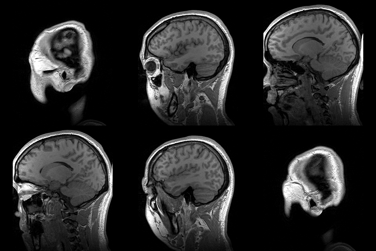
– An MRI (Magnetic Resonance Imaging) Scan is a painless imaging technique that uses magnets to take pictures of organs, muscles, and other tissues. Although an MRI may show a skull fracture, it takes better images of brain tissue, and areas of bleeding in or around the brain. Because MRI machines are very noisy and require a patient to lie still in a cramped space, sedation may be necessary. They also cannot be used for patients who have implanted metal devices such as pacemakers or knee replacements.
– An EEG (Electroencephalograph) is also a painless test where electrodes are attached to the scalp to measure the electrical activity of the brain. It is useful for patients who develop seizures as a result of their brain injury.
– An angiogram is a study of the blood vessels of the brain. A dye is injected into an artery that supplies blood to the brain. This test helps to determine if arteries or veins have been damaged, indicating a loss of blood supply to specific areas of the brain.
– An ICP (Intracranial Pressure) monitor is a tube placed through a small hole in the skull, and directly into or on the surface of the brain. A craniotomy is the procedure used drill the hole in the skull. Once placed, the ICP monitor measures the pressure of the cerebral spinal fluid. Any increased fluid within the skull cavity exerts pressure on the brain which causes pain and neurological impairment.
– The Glasgow Coma Scale (GCS) is an objective way to measure a patient’s level of consciousness after a traumatic brain injury. Three types of responses are assessed:
- Eye opening and wakefulness
- Motor responses (purposeful vs non-purposeful movements)
- Verbal responses (ability to talk, awareness of time, place and who they are)
The GSC is performed in the emergency room or after transfer to an intensive care unit. This scoring system is a way to rate symptoms, and determine what level of acute care is required. The lower the score, the more likely a severe brain injury has occurred.
Glasgow Coma Scale

“I learned more about brain injuries by talking to the doctors and especially the nurses who cared for my son. Most tried to be as honest as possible about the prognosis. The truth is, it’s a waiting game.”
– Mother of a 16- year-old who survived a two-week coma and a five-month hospital stay after a car accident.
The Glascow Coma Scale score is calculated by assigning a number to eye opening, verbal, and motor responses, then adding the three values together. Scores can range from 3 to 15 depending on the severity of brain injury. For example, if evaluating a patient’s motor responses, the ability to obey verbal commands is a “best response possible.” Patients who are on a ventilator for breathing support and cannot respond verbally may have a “t” notation with their score. In this scenario, the best possible score would be 11t. When there is no response to a verbal command, a painful stimulus is applied. Any response to pain indicates brain activity. Therefore, the GCS for a patient has died is 3.
The scales below represent the scores for children above age five, adolescents, and adults.
Glascow Coma Scale Test
Eye Response Score Patient Response
Spontaneously 4 eyes open spontaneously
To speech 3 eyes open to verbal commands
To pain 2 eyes open in response to pain
None 1 no eye opening in responses to stimulus
Motor Response
Obeys Commands 6 reacts to verbal commands
Localizes to Pain 5 identifies localized pain
Withdraws to Pain 4 flexes/withdraws from painful stimulus
Abnormal flexion 3 assumes a decorticate posture*
Abnormal extension 2 assumes a decerebrate posture*
None 1 no response or movement
Verbal Response
Oriented 5 fully aware and converses logically
Confused conversation 4 disoriented and confused
Inappropriate words 3 replies randomly with incorrect words
Incomprehensible sounds 2 moans or screams
None 1 no vocalizations or responses
Certain scores on the Glasgow Coma Scale have clinical significance. Patients with a score of 7 or less are considered comatose. Patients with a score of 8 or less are felt to have suffered a severe head injury.
*Decorticate Posture is the position of a comatose patient in which the arms are rigidly flexed at the elbows and wrists. The legs are extended with the toes rigidly pointed inward. After a head injury, this posture indicates bleeding on or within the brain, or increased intracranial pressure. In some instances, this posture may be produced by applying a painful stimulus to a comatose patient.
*Decerebrate Posture is the position of a comatose patient in which the arms are rigidly straight and hands are curled, while the legs are extended with the toes rigidly pointed downward. It is usually observed in patients experiencing compression of the lower brainstem.
 Brain Injury: A guide for family and friends
Table of Contents
• What is a Brain Injury?
• How Bad Is It?
• How the Brain Functions
• Common Problems During Early Recovery
• The Intensive Care Unit (ICU)
• Understanding Coma
• How Does an Injured Brain Heal?
• How You Can Help With Recovery
• Where Will the Journey Go From Here?
• How Will I Ever Get Through This?
• Where to Go for Help
• Books for Families Coping With Brain Injury
