Hand Bone X-Ray (Radiography) uses small amounts of electromagnetic radiation for imaging of bones in the hand and wrist. Bone X-Rays are painless, quick and simple ways of viewing bone and joint abnormalities for assessment, diagnosis and treatment purposes.
Hand X Ray: Definition
A hand x-ray is an imaging technique used to take pictures of hand bones using radiological waves known as “x” rays for medical purposes.
Who do I need to see to get an x ray?
If you have experienced an acute trauma to your hand, finger or wrist and think you may have an injury you should see your doctor. Similarly, if you have chronic pain, stiffness, swelling, weakness, numbness or tingling in your fingers, hand or wrist, you should see your doctor.
Your doctor may suggest that you need an x-ray to see what is going on with your hand, wrist and finger bones. The doctor will order for an x-ray image to be taken. A radiology technician will likely take the images and send them to a radiologist, or a specialist doctor trained to read and interpret x-ray images. The radiologist will then read the image and report the findings to your doctor, who will give the findings to you, the patient.
Cost of Hand X-Ray
The cost of a hand x-ray will depend on your location, type of insurance, reason for the x-ray, and if it is the whole hand, a finger, or the wrist.
Typically, one might expect to pay about $100 for a finger x-ray; $190 for a wrist x-ray; and $180 for a hand x-ray.
Imaging Techniques
X-rays are helpful in doing quick glimpses of bone issues such as breaks, and dislocations. For more detailed studies physicians may choose to use other types of imaging.
Some of the following other imaging techniques may be used with an x-ray for a more comprehensive examination:
Bone Scan
Bone scans are a type of imaging that uses small amounts of radioactive substance (injected into a vein and then settles into bones) to view absorption patterns into bones. The field of nuclear medicine uses this technique to diagnose metabolic activity in bones such as cancer, metastasis–or the spreading of cancer, fractures, infections or inflammation.
MRI
Magnetic Resonance Imaging is a type of imaging that uses magnetic energy to take images of internal structures including bones, organs and soft tissues such as muscles, tendons, nerves, and ligaments.
Ultrasound
Ultrasound imaging is a type of imaging that uses sound waves to capture images of internal structures of the human body. Ultrasound is unique in that it can allow for visualization of movement in real time, such as blood flow movement.
What can you see in an x ray?
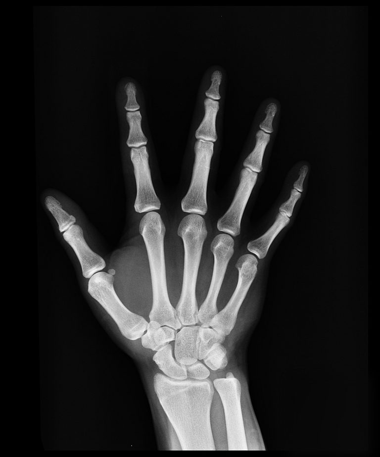
Normal Findings: Hand Bones/ Wrist Joint
In order to read and interpret x-ray imaging of the hand, one must be familiar with the normal anatomy of the hand.
Bones
The human hand is comprised of many small bones and joints that are a part of the skeletal system. Actually, the hand is made up of 27-29 different bones (some people may have more), 34 muscles, tendons and ligaments, nerves, arteries, veins and cartilage. There are a total of 14 joints present in the fingers and still more in the wrist.
The anatomical term for the hand and hand bones is carpal. (This is important in distinguishing bones of the feet from bones from the hand as they are labeled similarly. Feet bones are referred to as “tarsal.”)
The forearm bones that connect to the wrist are sometimes considered part of the hand and may be viewed in a hand x-ray. Those bones are known as the radius and ulna. The distal end of the radius bone connects to the wrist bones on the thumb side of the hand.
The small wrist bones are named the following:
- Scaphoid
- Lunate
- Triquetrum
- Pisiform
- Trapezium
- Trapezoid
- Capitate
- Hamate
Connected to the distal (away from the center of the body) end of the wrist bones are the five metacarpal bones, each forming part of the five fingers. Moving distally from the metacarpals, are the proximal phalanges, intermediate phalanges and then distal phalanges. This is the same in each of the fingers with the exception of the thumb, which does not have an intermediate phalange–only a proximal and distal phalange.
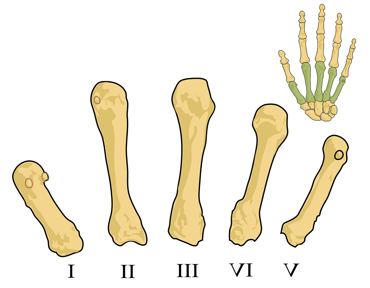
Metacarpals I-V
Abnormal Findings in Hand X-Ray
What might show up on a hand x ray?
Hands can be injured fairly easy as they are used for many different things. From jammed and severed fingers to breaks and carpal tunnel syndrome, the human hand is susceptible to may different types of acute and chronic injury of varying levels of severity.
If you are experiencing hand, wrist or finger pain, swelling, discoloration, numbness, tingling or weakness it is recommended to seek medical attention. A physician may perform a hand x-ray, MRI or ultrasound to rule out, assess, evaluate and diagnose the problem.
A hand x-ray is often used to determine type of injury, extent of injury, and helps to determine treatment of the injury. Hand x-rays can detect broken bones and arthritis of the hand.
Hand Injuries
Types of hand injuries may include but are not limited to: a cut (laceration); break (fractures); dislocation; muscle, tendon, ligament or cartilage damage; crushing injury; burn; or infection.
Cut (Laceration)
Lacerations to the hand can be very severe. There are many muscles, nerves and tendons in the hand and wrist that are susceptible to injury when a cut is experienced to the hand. Laceration damage to these soft tissues in the hand may result in immobility and/or loss of sensation. In other cases, a hand laceration may require some degree of amputation, and generates a high risk of excessive bleeding and infection.
Break (Fracture)
A break or fracture of the hand is when one or more of the bones of the hand has a crack in it or displays some degree of brokenness. There are different types of fractures and some of them are named based on their location or their cause. Examples include Boxer’s Fracture and Colles’ Fracture.
Boxer’s Fracture (also known as Brawler’s Fracture)
Boxer’s Fracture is a break or or more of the metacarpal bones (hand bones connecting the wrist to the phalanges). The classical definition of a Boxer’s Fracture is to the 4th or 5th metacarpal (ring finger or pinky), but some physicians consider a break to any of the metacarpals a Boxer’s Fracture. As its name suggests, the most common cause of a Boxer’s Fracture is when a closed fist punches a hard object.
Colles’ Fracture
Colles’ Fracture is a break of the distal radius bone of the forearm. It can be detected by a hand x-ray that includes the wrist and forearm.
Phalangeal Fractures
Of course the finger bones or phalanges (distal, intermediate and proximal) can also break. These breaks can be visualized by hand x-ray.
Crushing Injury
Crushing injuries are when a body part is subject to a forceful compression. These injuries can be very severe and can involve many different types of injuries such as fractures, dislocations, lacerations, contusions, foreign objects or object fragments imbedded into soft tissue, bleeding, nerve damage, tendon or ligament tearing, etcetera.
Treatment of hand, wrist or finger breaks
Sometimes a fracture can heal and be treated by stabilization and immobility of the joint by a cast or brace. In other cases, surgical implantation of a rod and screws may be necessary.
It is important to seek immediate medical attention if you think you have a broken bone in your wrist, hand or finger, as improper healing can cause chronic issues in the affected bone or joint such as stiffness, immobility, pain, numbness, tingling or weakness. People who leave these issues untreated may have difficulty performing normal daily tasks such as using a pen, using an eating utensil, buttoning, etcetera.
Joint Dislocation
Joint dislocation is when the bones of a joint are forced or otherwise moved out of their normal placement within the joint they make up. Dislocations are detectable by x-ray.
Risks of further injury with joint dislocation may include swelling, and tearing or damage to surrounding soft tissues such as cartilage, muscles, nerves, tendons or ligaments.
Muscle Damage
Typically muscle injury is not visualized in hand x-ray imaging, however, if hand pain is being diagnosed, an x-ray may be taken to rule out pain or discomfort of a broken bone or dislocated joint.
Strain (pulled muscle)
A muscle strain is likely the most common type of muscle injury in the hand. Simply a muscle strain is an overstretching of the muscle–a severe form of this could be a tearing (partial or complete) of the muscle.
Tendon and Ligament Damage
Ligament: Skier’s Thumb
Skier’s Thumb is an example of a hand ligament injury. Ligaments are soft tissue that connect bone to bone–in this case, the ulnar collateral ligament that connects the metacarpal to the proximal phalanx of the thumb is ruptured as a result of a sudden or forceful pulling of the thumb away from the other fingers of the hand. The rupture can be through the whole ligament or just partially through it.
Hand x-rays will not detect injury to ligaments because they are soft tissues. Injury to a ligament may need to be diagnosed via MRI imaging.
Sprain
Another type of ligament injury is known as a sprain. Sprains are in general, the injury to the soft tissue that connects bone to bone–ligaments. There are three degrees of sprains including an overstretching of the ligament, partial tear, and complete tear.
Sometimes when a sprain occurs the person hears a snap or pop sound. Symptoms of sprain include an unstable joint or loose/wobbly joint, swelling, pain, bruising, numbness, tingling, and/or a grating sound with movement.
Tendon: Mallet Finger
Mallet finger refers to injury or the rupture of a tendon near the fingertip. Injury to this tendon is usually caused by direct force to the fingertip (such as trying to catch a basketball, but the ball hits the fingertip and bends it awkwardly). Tendons connect muscle to bone, therefore injury to the tendon near the fingertip results in an inability to move or straighten the fingertip–it remains in a bent position.
Similarly to ligament injuries, an x-ray image will not detect injury to a tendon. MRI imaging is preferable to assessment and evaluation of soft tissue damage such as tendon injuries.
Nerve Damage
Carpal Tunnel Syndrome
The hand bone has several nerves that run to it helping to signal movement and sensation. One of the main nerves, called the median nerve, runs through the carpal bones of the wrist, in a “tunnel” known as the “carpal tunnel.” Overuse or repetitive movements (like typing) and arthritis of the wrist or hand can cause swelling and pressure to this nerve in the carpal tunnel resulting in a disorder known as Carpal Tunnel Syndrome.
An MRI, ultrasound or x-ray may be performed to examine if the pain, tenderness, numbness, weakness or tingling of the fingers, wrist and hand are due to Carpal Tunnel Syndrome or another cause.
Cartilage Damage
Arthritis
Arthritis is an example of cartilage damage that can be present in the hand. Arthritis of the bones in the hands can cause pain, stiffness, difficulty with movement, and be very uncomfortable. Arthritis may be visualized in hand x-ray imaging.
Hand x-ray findings for arthritis in the hand include the presence of osteophytes or periarticular erosion or the breaking down of bone around the joint.
- Osteophytes are a clinical symptom of osteoarthritis; they are bone spurs or boney growths that may be detected in x-ray imaging.
- Periarticular erosion is a clinical symptom of Rheumatoid Arthritis (RA). Cartilage and bone damage or destruction is the key sign of RA, an inflammatory disease.
Infection
Severe infection and inflammation of the bone and/or bone marrow, known as osteomyelitis can be detected, evaluated and assessed by x-ray imaging. In cases of osteomyelitis, a variety of imaging techniques may be used to assess other affected parts.
How is an X-Ray read/interpreted?
After an x-ray image is captured, it is sent to a radiologist to read and interpret.
The radiologist looks for discolorations, fractures, cracks or fissures, and abnormal growths. This information helps him/her to interpret the findings for the doctor. The doctor takes the information and puts it together with other signs and symptoms and assessment findings to make a diagnosis.
Preparation for hand x-ray
If you are able to remove any jewelry, rings, bracelets or watches from your hand and wrist before an x-ray this is preferred. However, if the injury you have sustained does not allow for the removal of accessories, an x-ray can be taken with these accessories present.
Risks of x-ray imaging
Risks of x-ray imaging of the finger, wrist or hand are very low. In general for any x-ray imaging there are two main risks: 1) development of cancer and 2) risk to fetus in utero.
The amount and exposure to radiation in a hand x-ray is very low, therefore the risk of developing cancer from a hand x-ray is very low.
In addition, women who think they may be pregnant, are pregnant, or are planning to become pregnant are asked to wear a lead apron to protect their abdomen and reproductive organs. Children will also be asked to wear protective aprons to protect their reproductive organs.
