X-ray of the knee, otherwise known as startradiology, is the use of small amounts of radiation to capture an image of the bones in and around the knee joint.
Bone X-Ray: Definition
Bone X-Ray (Radiography) uses small amounts of electromagnetic radiation for imaging of any bone in the human body. Bone X-Rays are painless, quick and simple ways of viewing bone or joint abnormalities for examination, assessment and diagnosis.
This article discusses the use of bone X-Ray for problems in the knee.
Other types of imaging
Bone Scan
Bone scans are typically performed in the medical field known as nuclear medicine. It is an imaging technique that is used to view different types of bone problems. A small amount of a radioactive substance is injected into a vein and settles into bones. Depending on the absorption of this tracing substance, nuclear medicine specialists can diagnose abnormal metabolic activity in bones (referring to cancer, the spreading of cancer (metastasis), fractures, infection or inflammation).
Magnetic Resonance Imaging
Magnetic Resonance Imaging (MRI) is a technique used to capture detailed images of the inside of the human body. It uses magnetic forces, radio waves and computer technology to take pictures of the body’s organs, bones or soft tissues. A big difference between MRI and X-Ray is that an MRI does not use radiation.
Magnetic Resonance Enterography
Magnetic Resonance Enterography (MRE) is a type of MRI that allows for imaging of the inside of the small intestine or bowels.
Magnetic Resonance Angiography
Magnetic Resonance Angiography (MRA) is a type of MRI that allows for examination of blood vessels.
Ultrasound Imaging
An ultrasound uses high-frequency sound waves (or ultrasound) to capture images of internal body organs, muscles, tendons, joints and blood vessels. A unique feature of ultrasound technology is that it can capture movement of internal structures, including blood flow, and it can be used to view structures in real time.
Computed Tomography Scan
Computed Tomography Scan (CT Scan or CAT Scan) is an imaging technique that takes a series, or combination, of x-ray images of a certain part of the body. These many images are then stacked together to form a three-dimensional image of the organ, joint, tumor or other internal structure of interest.
Who do I need to see to get an x-ray?
If you think you might need an x-ray, likely you should first see your primary care physician. They will then direct you to a radiologist, or a specialist doctor trained to read and interpret x-ray images. The radiologist may or may not be the person who actually takes the images, but likely they will read the image and report the findings to your doctor, who will then give the findings to you, the patient.
How is an X-Ray read/interpreted?
After an x-ray image is captured, it is sent to a radiologist to read and interpret. The radiologist must know what a normal healthy knee looks like so that she/he can identify problems.
The radiologist looks for discolorations, fractures, cracks or fissures, abnormal growths, and takes measurements of the space between bones. This information helps him/her to interpret the findings for the doctor. The doctor takes the information and puts it together with other signs and symptoms and assessment findings to make a diagnosis.
Purpose of X-Ray
The purpose of x-ray imaging is to obtain a quick image of bone for assessment, evaluation, and/or diagnosis of bone disorders such as fractures, dislocations, cancers, infections, osteoarthritis, and abnormal bone growths.
The following presents a list of the different uses and purposes of a bone x-ray.
Diagnose
- Bone fractures or breaks
- Dislocation of joint
- Bone cancer
- Locate
Locate
- X-Ray images may be used to help locate presence of foreign objects nearby bones.
- After a surgery, a surgeon may request an x-ray to detect the presence (or confirm the absence) of any tools or materials that may have been left inside the patient.
Assess
- After treatment of a bone fracture or break, an x-ray image may be taken to ensure that the bone and/or joint has healed properly (alignment, posture, no loose bone fragments).
- During surgery an x-ray image may be used to view bone repairs, fusions, replacements or fracture reductions.
Evaluate
- In certain metabolic disorders, x-ray imaging may be used to evaluate changes in abnormal bone growth, changes in arthritis, infection or injury.
Risks of x-ray imaging
The two main risks associated with x-ray imaging are 1) development of cancer and 2) risk to fetus in utero.
Cancer Risk
The risk of developing cancer due to radiation exposure from x-ray imaging, while present, is fairly low. In today’s technology, scientists and physicians have developed methods for using very controlled radiation beams that do not scatter much into other organs and methods for using the lowest dose of radiation necessary for the imaging.
Pregnancy and X-Ray
Always tell your doctor or medical staff if you think you might be pregnant or are planning to become pregnant before having an x-ray.
Having a large number of x-rays to the abdominal region before awareness or knowledge of pregnancy may cause miscarriage.
It is possible to have x-ray imaging while pregnant. Likely if the x-ray is done on an arm or leg, there poses very minimal risk to the fetus, the mother will still be asked however to wear a lead apron to protect the abdomen and fetus from scattered radiation beams.
If the x-ray is needed of the abdominal region, your physician will discuss the risks and benefits with you and help you to determine the best decision for you and your baby. The risks posed to the baby depend on the gestational age of the baby.
Positioning required for knee x-ray
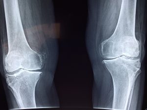
AP
The anterior to posterior (AP) view is the standard front-to-back view of the knee joint. The patient is either supine (lying face up) or standing, and the image is taken of the knee from the anterior (front) side.
PA
The posterior to anterior (PA) view (also referred to as Rosemberg method), captures an image from the back-to-front of the knee joint. The patient is standing with their knees bent at a 45 degree angle. The image is taken from the posterior (back) side.
Merchant View
Merchant view, or Skyline Merchant View, refers to the position of the x-ray image taken of the knee, where the patient is supine (face up) with their feet hanging off the edge of the exam table at a 45 degree angle. In this particular image, primarily the patella is viewed. An image in this position can be useful in determining: the patellar tilt, patellofemoral arthritis, and fracture of the patella.
Knutsson View
Similarly to the Merchant view, the Knutsson view requires the patient to be in a supine (face up) position but their knees are at an angle with their feet on top of the exam table.
Sunrise View
Sunrise or Settegest view of the knee, refers to the position of the x-ray image where the patient is prone (face down) with their knees bent at an acute angle (bottom of the feet point toward the head). The imaging equipment (camera part) suspended above and is perpendicular with the patient’s knee cap. When the image is printed, the kneecap appears as a sunrise with the joint.
Jaroschy
Similarly to the Settegest view, the Jaroschy view requires the patient to be in a prone (face down) position on the exam table. The patient’s knees are bent at an obtuse angle (toes point away from the head).
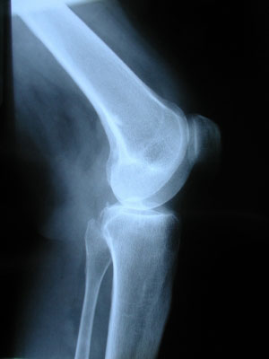
Lateral View
The lateral view is a viewing of the side of the knee. The patient lies supine with their knee bent at a 30 degree angle and the x-ray waves pass through the medial (inner) side to the lateral (outer) side.
There are many other variations and positions for knee x-ray images to be taken. The position and angles required depend on the reason for the x-ray imaging and the trauma or injury the patient is experiencing (ability to get into a certain position). Sometimes, physician may want several images of the knee in a variety of positions and angles to determine different measurements.
Cost of knee x-ray
Of course the cost of a knee x-ray will depend on a variety of factors such as location, reason for x-ray, insurance, etcetera; however, a an estimation of $200 per image may be expected.
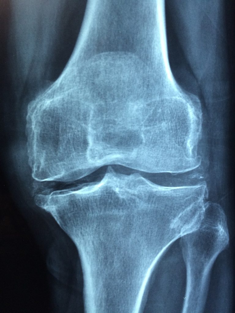
What can you see in a knee x-ray?
Typically, in a standard knee x-ray, the knee joint bones are primarily viewed; while as ligaments, tendons and meniscus are not easily viewed.
In order to view ligaments, tendons or meniscus a CT scan or MRI will be needed.
Normal Findings: Knee Bones/ Medical Knee
The knee is a synovial joint, meaning it allows for movement. The knee is also considered a hinge joint (synovial hinge joint), which means it allows for bending and straightening movements.
Bones
- Patella- kneecap
- Tibia- shin bone
- Femur- thigh bone
- *Fibula- technically this is not considered part of the knee or knee joint but can show up in knee x-ray images. It is the bone on the lateral (outer) side of the lower leg–or calf bone.
Knee Joint
The knee actually has two joints known as articulations:
- Patellofemoral Articulation- made up by the patella (kneecap) and femur bone
- Tibiofemoral Articulation- hinge joint made up by the tibia bone and femur bone
Ligaments
Ligaments connect bone to bone. In the knee, ligaments are named based on their positions in the knee joint.
- Anterior Cruciate Ligament (ACL)
- Posterior Cruciate Ligament (PCL)
- Medial Collateral Ligament (MCL)
- Lateral Collateral Ligament (LCL)
- Patellar Ligament
Tendons
Tendons connect muscle to bone. The main tendon in the knee joint is called the patellar tendon. It connects the quadriceps muscle (thigh muscle) to the patella.
Meniscus
Two meniscus, or shock-absorbing discs made of connective tissue, cushion the condylar ends of the femur bone and the tibia bone. The menisci are named for their locations in the knee joint–medial (inner) and lateral (outer).
Abnormal Findings
What might show up on a knee x ray?
- Osteoarthritis
- Signs of extra fluid and swelling
- Fracture
- Abnormal bone structure
- Abnormal alignment of the knee joint
Findings of Osteoarthritis in Knee X-Ray Imaging
- On an x-ray, cartilage cannot be assessed, but the space between bones decreases significantly in cases of knee arthritis. This condition is known as joint space narrowing and can be measured in a knee x-ray. Knee joint space narrowing is a clinical sign of arthritis of the knee.
- Osteophytes, or bone spurs, may also be detected in knee x-ray imaging. These abnormal bone growths are symptomatic and signs of osteoarthritis.
Findings of Fractures in Knee X-Ray Imaging
While not all fractures can be visualized on x-ray imaging, some can! They may appear as a dark thin line or crack through the white bone. In severe bone breaks you may see the bone completely separated by a thick dark space.
Common fractures to visualize in a knee x-ray are:
- Tibial plateau fracture
- Patella fracture
Findings of Abnormal Bone Structure in Knee X-Ray Imaging
- Bone thinning (osteopenia) can be measured and detected in an x-ray of the knee. Osteopenia is a structural abnormality of the bone.
Preparation for knee x ray
There is not much preparation needed for a knee x-ray. Simply, show up to the doctor’s office or hospital. They will give you instructions to remove jewelry or possibly some clothing, perhaps wear a hospital gown, and to position yourself a certain way.
Do make sure that if you are pregnant or planning to become pregnant that this is communicated clearly to your medical team.
If you have had any problems with x-ray or other radiation in the past make sure to also communicate this with your medical team.
Equipment
Typically, a large “camera” is attached to a large arm that can be moved easily to take pictures from many different angles. A smaller board will be used to provide a “back” or background for the image. You may asked to hold it at a certain angle or to rest your knee on top of it.
You may also be asked to wear a lead apron over your chest and abdomen, especially if you are pregnant or planning to become pregnant. This simply helps to prevent any scattered x-rays to travel to your chest and abdominal areas.
What to expect
You may wonder what to expect if you are going in for a knee x-ray. Having an x-ray of the knee is similar to getting pictures taken of your knee. It is not painful–however if you have a knee injury the positioning and need to ‘hold still’ in that position may be uncomfortable.
Likely you will either need to stand, sit or lay down and bend your knee or knees in a certain way. The technician, nurse or doctor will help you to get into the right position. Then, you will need to hold still while they position the “camera.”
What to wear
If you need to have your knees x-rayed it is probably a good idea to wear a skirt or shorts so that your knees are visible. You may be asked to change into a hospital gown for the x-ray image.
You may also be asked to remove your bra for an x-ray. If you prefer to keep it on, wear a sports bra with no wires or metal clasps.
It is important not to wear metal or jewelry during an x-ray. Tell your doctor if you have any implants or pacemaker.
