Statistics have shown that plantar fasciitis (PF) is the most common reason for heel pain. This condition results from micro-trauma to the plantar fascia – an elongated stretch of tissue that supports the arches under the feet. However, recent studies suggest that many cases of plantar fasciitis are actually misdiagnosed.
Parading as plantar fasciitis, Baxter’s Neuropathy very closely resembles the clinical picture for PF. However, because there isn’t a definitive diagnostic test for Baxter’s Nueropathy, most physicians choose to diagnose majority of heel pain complaints as plantar fasciitis. A revision of these diagnoses is often made after treatments for PF have been administered but failed to provide substantial improvement.
The problem here is that treatments for plantar fasciitis can be varied. Some of these methods may even be detrimental to Baxter’s Nueropathy. So it’s imperative to rule out plantar fasciitis at the onset of the condition to be able to provide appropriate treatment that won’t exacerbate your pain.
The question now is – how can you be certain that your heel pain is the result of Baxter’s Neuropathy? To better answer that, I’ve come up with a definitive, in-depth discussion on Baxter’s Neuropathy, its origins, diagnosis, and treatment.
A Quick Lesson in Foot Anatomy
A large part of understanding Baxter’s Neuropathy relies on the knowledge of the normal foot anatomy. Internal structures have a great impact on the development and progression of Baxter’s Neuropathy. So, a clear picture of the anatomy inside the foot can help provide practical information on the proper handling techniques during various types of treatment.
Baxter’s Neuropathy is a condition affecting Baxter’s Nerve, or the inferior calcaneal nerve (ICN). This particular nerve supplies sensory functions to the areas of the plantar surface of the foot, and motor function to several muscles including the abductor digiti minimi and the flexor digitorum.
The location and structure of the inferior calcaneal nerve is largely responsible for its susceptibility to impingement. It starts off from the tibial nerve, which branches out as it passes through the tarsal tunnel. Here, along the medial side of the ankle, the two branches – the medial and lateral plantar nerve – make a sharp turn to innervate structures underneath the foot.
The lateral branch is that which gives rise to the inferior calcaneal nerve. This small projection can be found just under the medial surface of the foot. But in some people, the ICN can be found much more proximally. There have been some cases when the ICN branched out as early as the ankle, just around the tarsal tunnel.
How is the ICN Involved in Impingement?
The location of the ICN is clinically significant because it plays a role in the nerve’s susceptibility to impingement. The tight turn the branch takes as it moves from the arch of the foot to innervate the structures underneath make it especially prone to the tensile and compressive forces of an individual’s weight.
Most commonly, the ICN is obstructed in one of two ways. The first is the between the quadratus plantae muscle and the fascia of deep fascia of the abductor hallucis. The second most likely location for impingement is along the medial calcaneal tuberosity, especially if an individual has developed bone spurs in this region.
Depending on the location and structure of a person’s ICN, they may be more or less prone to ICN impingement. Usually, those whose ICN branches out more distally are more susceptible to this type of neuropathy.
Diagnosing Baxter’s Neuropathy
There’s very little literature on Baxter’s Neuropathy, and it’s often overlooked or ruled out by doctors because there’s no way to truly confirm its presence other than with an MRI. Because most patients would rather not go through the process of such a tedious imaging test just for pain in their feet, many cases go falsely diagnosed for months.
However, there are certain methods that doctors and perhaps even you can perform in order to verify whether or not Baxter’s Neuropathy is present. The first is by taking the history of the individual into consideration.
There are several factors that could predispose a person to Baxter’s Neuropathy. These include:
- Obesity
- Sudden increase in running distance
- Occupations that require long periods of standing or walking
- Flat footedness
- Plantar fasciitis
- Limitation of ankle range of motion
All of these factors somehow contribute to the likelihood of the ICN becoming impinged between surrounding structures. These alter the biometrics of the foot, and thus increase the tensile and compression forces throughout the structures.
If an individual possesses a combination of these predisposing factors, then it would be practical to assess for Baxter’s Neuropathy. There are certain physical tests that a medical practitioner can perform to determine the presence of ICN impingement. However, it’s important to note that these tests can only be definitive if they’re interpreted in combination with MRI findings.
Palpation and Inspection
Because the ICN is a mixed motor-sensory nerve, it provides sensation throughout different areas of the plantar surface of the feet. In the involved side, ICN can cause significant pain and pronounced numbness. This is usually most evident along the length of the arch of the feet, as well as the lateral side where the abductor digiti minimi is located.

While it isn’t as easy for the untrained eye, it’s also possible for a specialist to detect subtle differences in the bulk of the muscles affected by the impingement. Obstruction of the motor function of the ICN can result to atrophy of its innervated muscles in the long run, so comparing the affected foot to the unaffected will potentially reveal differences in muscle mass.
Testing Muscle Strength
Another way of determining the presence of Baxter’s Neuropathy is by checking the strength of the muscles involved. The ICN innervates the flexor digitorum muscle, which works to flex the four smaller toes. When the nervous supply to this muscle is limited or obstructed, the toes may lose strength during resistive flexion.
To test this, ask the individual to step on a small card. Instruct them to prevent the card from being pulled from under their toes. Then gently and slowly pull the card away. If the individual struggles to resist, or if the card can be easily pulled with minimal resistance, then Baxter’s Neuropathy should be considered.
If you’re having a hard time interpreting the results, try performing the same test on the unaffected foot. A stronger resistive force could indicate unilateral weakness caused by ICN impingement.
MRI Scan
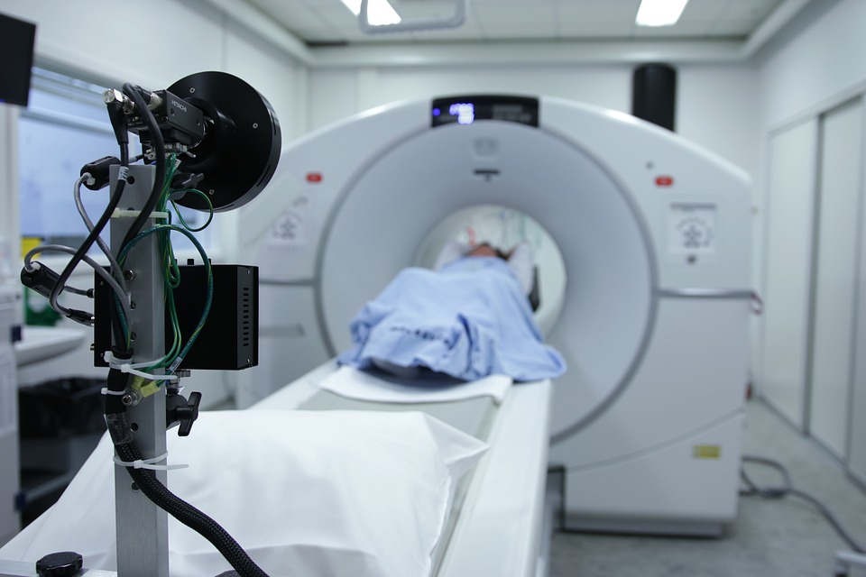
The most reliable test for Baxter’s Neuropathy is an MRI scan. This imaging test can reveal the accumulation of fatty edema, as well as the likely atrophy of the involved muscles. With this imaging test, most doctors can rule out any other differential diagnosis and thus confirm the presence of Baxter’s Neuropathy.
However, since very few individuals will opt to undergo an MRI for something as common as foot pain, ICN impingement will often go undiagnosed. In majority of cases, doctors will claim that the symptoms point to plantar fasciitis.

The problem here is that because the treatments for plantar faciitis and Baxter’s Neuropathy are very different, it’s possible to exacerbate the symptoms of the condition and potentially compound consequent complications. So it’s suggested to consider Baxter’s Neuropathy if foot pain becomes chronic and traditional methods of treatment fail to relieve the symptoms.
Treatments for Baxter’s Neuropathy
Initial management for Baxter’s Neuropathy is conservative. Some doctors will recommend that individuals seek the assistance of physical therapists to manipulate the soft tissues and release tensile forces. A clear understanding of human anatomy is necessary for anyone who attempts to manually relieve the pain as manipulation of muscles and soft tissues in the wrong direction could lead to further complications.

Commonly, the most effective movement for lessening pain is inversion of the ankle. The inward movement of the foot reduces tension on the ICN significantly, thus relieving pain temporarily during treatment. Repetitive performance of such manipulation should help to reduce pain over time, and may help the brain recalculate sensation to further decrease the intensity of discomfort.
Other treatments for Baxter’s Neuropathy include rest, corticosteroid injections, NSAIDs, and orthotics. However, if pain sustains or worsens over time, then doctors might recommend operative intervention.
During the procedure, surgeons will remove or nip away portions of plantar fascia to relieve compression. If deep fascia connected to the abductor hallucis is also involved, then doctors will also release a portion of it to relieve tensile forces on the ICN.
Complications are highly unlikely after such a procedure, and a full recovery can be expected within a few weeks of the surgery.
Conclusion
Historically, Baxter’s Neuropathy rarely progresses to more serious complications. However, pain may increase significantly as time wears on, so anyone who suffers from the condition may experience grave changes in occupational and social participation.
With that, it’s important to make sure you consider all the angles once you experience those first few episodes of foot pain. Keep in mind that Baxter’s Neuropathy isn’t always considered a primary diagnosis. So it’s important to collect all the facts of your condition before ruling it out to prevent the administration of treatments that could potentially worsen the case.
In the event that Baxter’s Neuropathy is confirmed, conservative treatments are a far better solution to try first before you undergo any sort of surgery. This is especially true if pain is tolerable enough. However, it’s of utmost importance to perform any management under the supervision and guidance of a licensed practitioner, as home remedies can cause further complications.